Mice lacking monocyte chemoattractant protein 1 have enhanced susceptibility to an interstitial polymicrobial infection due to impaired monocyte recruitment
- PMID: 12011011
- PMCID: PMC127982
- DOI: 10.1128/IAI.70.6.3164-3169.2002
Mice lacking monocyte chemoattractant protein 1 have enhanced susceptibility to an interstitial polymicrobial infection due to impaired monocyte recruitment
Abstract
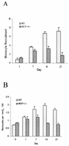
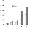
Monocyte chemoattractant protein 1 (MCP-1) is an important chemokine that induces monocyte recruitment in a number of different pathologies, including infection. To investigate the role of MCP-1 in protecting a host from a chronic interstitial polymicrobial infection, dental pulps of MCP-1(-/-) mice and controls were inoculated with six different oral pathogens. In this model the recruitment of leukocytes and the impact of a genetic deletion on the susceptibility to infection can be accurately assessed by measuring the progression of soft tissue necrosis and osteolytic lesion formation. The absence of MCP-1 significantly impaired the recruitment of monocytes, which at later time points was threefold higher in the wild-type mice than in MCP-1(-/-) mice (P < 0.05). The consequence was significantly enhanced rates of soft tissue necrosis and bone resorption (P < 0.05). We also determined that the MCP-1(-/-) mice were able to recruit polymorphonuclear leukocytes (PMNs) to a similar or greater extent as controls and to produce equivalent levels of Porphyromonas gingivalis-specific total immunoglobulin G (IgG) and IgG1. These results point to the importance of MCP-1 expression and monocyte recruitment in antibacterial defense and demonstrate that antibacterial defense is not due to an indirect effect on PMN recruitment or modulation of the adaptive immune response.
Figures


Similar articles
-
Immune response to Fusobacterium nucleatum and Prevotella intermedia in patients with peritonsillar cellulitis and abscess.Clin Infect Dis. 1995 Jun;20 Suppl 2:S220-1. doi: 10.1093/clinids/20.supplement_2.s220. Clin Infect Dis. 1995. PMID: 7548558 No abstract available.
-
Host modulation of tissue destruction caused by periodontopathogens: effects on a mixed microbial infection composed of Porphyromonas gingivalis and Fusobacterium nucleatum.Microb Pathog. 1997 Jul;23(1):23-32. doi: 10.1006/mpat.1996.0129. Microb Pathog. 1997. PMID: 9250777
-
Interleukin-1 receptor signaling rather than that of tumor necrosis factor is critical in protecting the host from the severe consequences of a polymicrobe anaerobic infection.Infect Immun. 2000 Aug;68(8):4746-51. doi: 10.1128/IAI.68.8.4746-4751.2000. Infect Immun. 2000. PMID: 10899881 Free PMC article.
-
The production of monocyte chemoattractant protein-1 (MCP-1)/CCL2 in tumor microenvironments.Cytokine. 2017 Oct;98:71-78. doi: 10.1016/j.cyto.2017.02.001. Epub 2017 Feb 8. Cytokine. 2017. PMID: 28189389 Review.
-
Immunological Pathways Triggered by Porphyromonas gingivalis and Fusobacterium nucleatum: Therapeutic Possibilities?Mediators Inflamm. 2019 Jun 24;2019:7241312. doi: 10.1155/2019/7241312. eCollection 2019. Mediators Inflamm. 2019. PMID: 31341421 Free PMC article. Review.
Cited by
-
Microarray analysis of the effect of Streptococcus equi subsp. zooepidemicus M-like protein in infecting porcine pulmonary alveolar macrophage.PLoS One. 2012;7(5):e36452. doi: 10.1371/journal.pone.0036452. Epub 2012 May 2. PLoS One. 2012. PMID: 22567158 Free PMC article.
-
MCP-1 deficiency delays regression of pathologic retinal neovascularization in a model of ischemic retinopathy.Invest Ophthalmol Vis Sci. 2008 Sep;49(9):4195-202. doi: 10.1167/iovs.07-1491. Epub 2008 May 16. Invest Ophthalmol Vis Sci. 2008. PMID: 18487365 Free PMC article.
-
FOXO1 deletion reduces dendritic cell function and enhances susceptibility to periodontitis.Am J Pathol. 2015 Apr;185(4):1085-93. doi: 10.1016/j.ajpath.2014.12.006. Am J Pathol. 2015. PMID: 25794707 Free PMC article.
-
Immunization enhances inflammation and tissue destruction in response to Porphyromonas gingivalis.Infect Immun. 2006 Apr;74(4):2286-92. doi: 10.1128/IAI.74.4.2286-2292.2006. Infect Immun. 2006. PMID: 16552059 Free PMC article.
-
Review of osteoimmunology and the host response in endodontic and periodontal lesions.J Oral Microbiol. 2011 Jan 17;3. doi: 10.3402/jom.v3i0.5304. J Oral Microbiol. 2011. PMID: 21547019 Free PMC article.
References
-
- Alam, R., J. York, M. Boyars, S. Stafford, J. Grant, J. Lee, P. Forsythe, T. Sim, and N. Ida. 1996. Increased MCP-1, RANTES, and MIP-1alpha in bronchoalveolar lavage fluid of allergic asthmatic patients. Am. J. Respir. Crit. Care Med. 153:1398-1404. - PubMed
-
- Bossink, A., L. Paemen, P. Jansen, C. Hack, L. Thijs, and J. Van Damme. 1995. Plasma levels of the chemokines monocyte chemotactic proteins-1 and -2 are elevated in human sepsis. Blood 86:3841-3847. - PubMed
Publication types
MeSH terms
Substances
Grants and funding
LinkOut - more resources
Full Text Sources
Molecular Biology Databases
Research Materials
Miscellaneous

