. 2002 Jun;70(6):3199-207.
doi: 10.1128/IAI.70.6.3199-3207.2002.

Apoptosis in acute shigellosis is associated with increased production of Fas/Fas ligand, perforin, caspase-1, and caspase-3 but reduced production of Bcl-2 and interleukin-2

Affiliations

Apoptosis in acute shigellosis is associated with increased production of Fas/Fas ligand, perforin, caspase-1, and caspase-3 but reduced production of Bcl-2 and interleukin-2

Rubhana Raqib et al. Infect Immun. 2002 Jun.

Abstract

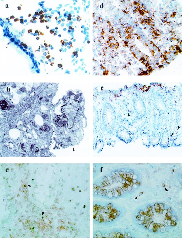
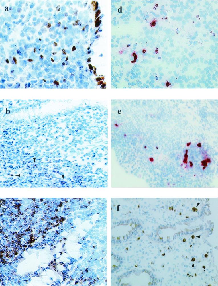
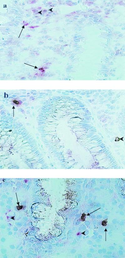
Shigella dysenteriae type 1-induced apoptotic cell death in rectal tissues from patients infected with Shigella dysenteriae type 1 was studied by the terminal deoxynucleotidyltransferase-mediated dUTP-biotin nick end labeling (TUNEL) technique and annexin V staining. Expression of proteins and cytokines participating in the apoptotic process (caspase-1, caspase-3, Fas [CD95], Fas ligand [Fas-L], perforin, granzyme A, Bax, WAF-1, Bcl-2, interleukin-2 [IL-2], IL-18, and granulocyte-macrophage colony-stimulating factor) in tissue in the acute and convalescent stages of dysentery was quantified at the single-cell level by in situ immunostaining. Apoptotic cell death in the lamina propria was markedly up-regulated at the acute stage (P < 0.05), where an increased number of necrotic cells were also seen. Phenotypic analysis of apoptotic cells revealed that 43% of T cells (CD3), 10% of granulocytes (CD15), and 5% of macrophages (CD56) underwent apoptosis. Increased activity of caspase-1 persisted in the rectum up to 1 month after onset. More-extensive expression of Fas, Fas-L, perforin, caspase-3, and IL-18, but not IL-2, at the acute stage than at the convalescent stage was observed. Increased expression of caspase-3 and IL-18 in tissues with severe inflammation compared to expression in those with mild inflammation was evident, implying a possible role in the perpetuation of inflammation. Significantly reduced cell death during convalescence was associated with a significant up-regulation of Bcl-2, Bax, and WAF-1 expression in the rectum compared to that in the acute phase of infection. Thus, induction of apoptosis at the local site in the early phase of S. dysenteriae type 1 infection was associated with a significant up-regulation of Fas/Fas-L and perforin and granzyme A expression and a down-regulation of Bcl-2 and IL-2, which promote cell survival.


Figures

FIG. 1.
FIG. 1.
In acute shigellosis, notable increases in apoptotic T cells, granulocytes, and macrophages, along with an increased number of necrotic cells in the rectal mucosa, are seen. A significant up-regulation of noncaspase regulators of apoptosis (Fas/Fas-L and perforin and granzyme A expression) is also evident. (a) Immunohistochemistry of the rectal mucosa of a patient with Shigella infection. Visible is extensive expression of TUNEL-positive cells (brown) and debris in the lamina propria during the acute stage of shigellosis. Magnification, ×320. (b) Electron micrograph of a rectal-surface epithelial cell (arrowhead) from an adult patient with shigellosis showing focal necrosis. Evidence of degeneration includes loss of microvilli, dilated rough endoplasmic reticulum, vesiculation, large autolysosomes, and nuclear changes. Magnification, ×5,440. (c) Double immunostaining showing TUNEL-positive (green; arrowheads) CD3 T cells (brown) in the lamina propria in a rectal biopsy sample from an acute Shigella-infected patient. Magnification, ×320. (d) Extensive expression of CD95 (Fas) in the lamina propria and in the epithelial lining of the rectal mucosa in patients during the acute stage of shigellosis. Magnification, ×320. (e) Expression of Fas-L in cells mostly located close to the surface epithelium, with some scattered in the deeper lamina propria (arrowheads), in rectal biopsy samples from a patient with shigellosis. Magnification, ×80. (f) Perforin-expressing CD8+ lymphocytes in the lamina propria in the rectum during the acute stage of Shigella infection. Arrowheads, double-positive cells (brown and pink). Magnification, ×320.
FIG. 2.
FIG. 2.
During acute Shigella infection, there was up-regulation of proapoptotic enzymes (caspase-1 and caspase-3) and substrate IL-18 and a down-regulation of an antiapoptotic marker (Bcl-2) in the local site of infection. During convalescence, there was increased expression of antiapoptotic protein Bcl-2 in parallel with a reduction in cell death in the rectum. (a) Expression of WAF-1 in tissue was localized mainly to the cells in the epithelium and some cells in the lamina propria in the rectum during the course of shigellosis. The staining pattern was cytoplasmic. Magnification, ×320. (b) Expression of the membrane-bound form of Bcl-2 was markedly reduced in the rectum in the acute stage of shigellosis. There were few Bcl-2-expressing cells present in the lamina propria close to the crypts (arrowheads). Magnification, ×320. (c) The expression of Bcl-2 in tissue increased considerably during recovery from acute shigellosis, with massive numbers of cells expressing the Bcl-2 protein. Magnification, ×320. (d) Double immunohistochemical staining showing apoptotic (TUNEL-positive, brown) caspase-3 (red)-expressing cells in the lymphoid aggregates. Magnification, ×320. (e) Apoptotic cells (TUNEL-positive, brown) expressing caspase-1 (red) were localized in the same lymphoid aggregate as that seen in Fig. 3a. Magnification, ×320. (f) IL-18-expressing cells (arrowheads) infiltrating the rectal mucosa in acute shigellosis. Mostly, IL-18-expressing cells were in close proximity to eosinophils. Magnification, ×320.
FIG. 3.
FIG. 3.
Various inflammatory cells infiltrate the local site of infection in the rectum during acute shigellosis and express proapoptotic molecules. (a) Fas-L-expressing CD68+ macrophages (deep pink and brown; arrows) were seen in close proximity to the luminal crypts in the lamina propria. Some Fas-L-expressing cells (brown only; arrowheads) that did not stain for CD68+ macrophages were also present in the vicinity. (b) Perforin-expressing CD8+ T lymphocyte (arrow) in an inflamed rectum during the acute stage of Shigella infection. A single perforin-expressing cell (arrowhead) close to a crypt was also seen. (c) A number of CD15-expressing granulocytes that coexpressed proapoptotic enzyme caspase-1 (arrows) were present in the rectal tissue during the early stage of shigellosis. Some other cell types that also expressed caspase-1 (single staining, pink; arrowheads) were seen near the double-positive granulocytes.

Similar articles

Cited by

References

    1. Akbar, A. N., and M. Salmon. 1997. Cellular environments and apoptosis: tissue microenvironments control activated T-cell death. Immunol. Today 18:72-76. - PubMed
    1. Aliprantis, A. O., R. B. Yang, M. R. Mark, S. Suggett, B. Devaux, J. D. Radolf, G. R. Klimpel, P. Godowski, and A. Zychlinsky. 1999. Cell activation and apoptosis by bacterial lipoproteins through toll-like receptor-2. Science 285:736-739. - PubMed
    1. Brach, M. A., S. deVos, H. J. Gruss, and F. Herrmann. 1992. Prolongation of survival of human polymorphonuclear neutrophils by granulocyte-macrophage colony-stimulating factor is caused by inhibition of programmed cell death. Blood 80:2920-2924. - PubMed
    1. Clerc, P., B. Baudry, and P. J. Sansonetti. 1988. Molecular mechanisms of entry, intracellular multiplication and killing of host cells by shigellae. Curr. Top. Microbiol. Immunol. 138:3-13. - PubMed
    1. Cohen, A., V. Madrid-Marina, Z. Estrov, M. H. Freedman, C. A. Lingwood, and H. M. Dosch. 1990. Expression of glycolipid receptors to Shiga-like toxin on human B lymphocytes: a mechanism for the failure of long-lived antibody response to dysenteric disease. Int. Immunol. 2:1-8. - PubMed

Publication types

MeSH terms

Substances