. 2002 May 13;157(4):591-602.
doi: 10.1083/jcb.200202047. Epub 2002 May 13.

The kinetically dominant assembly pathway for centrosomal asters in Caenorhabditis elegans is gamma-tubulin dependent

Affiliations

The kinetically dominant assembly pathway for centrosomal asters in Caenorhabditis elegans is gamma-tubulin dependent

Eva Hannak et al. J Cell Biol. .

Abstract

gamma-Tubulin-containing complexes are thought to nucleate and anchor centrosomal microtubules (MTs). Surprisingly, a recent study (Strome, S., J. Powers, M. Dunn, K. Reese, C.J. Malone, J. White, G. Seydoux, and W. Saxton. Mol. Biol. Cell. 12:1751-1764) showed that centrosomal asters form in Caenorhabditis elegans embryos depleted of gamma-tubulin by RNA-mediated interference (RNAi). Here, we investigate the nucleation and organization of centrosomal MT asters in C. elegans embryos severely compromised for gamma-tubulin function. We characterize embryos depleted of approximately 98% centrosomal gamma-tubulin by RNAi, embryos expressing a mutant form of gamma-tubulin, and embryos depleted of a gamma-tubulin-associated protein, CeGrip-1. In all cases, centrosomal asters fail to form during interphase but assemble as embryos enter mitosis. The formation of these mitotic asters does not require ZYG-9, a centrosomal MT-associated protein, or cytoplasmic dynein, a minus end-directed motor that contributes to self-organization of mitotic asters in other organisms. By kinetically monitoring MT regrowth from cold-treated mitotic centrosomes in vivo, we show that centrosomal nucleating activity is severely compromised by gamma-tubulin depletion. Thus, although unknown mechanisms can support partial assembly of mitotic centrosomal asters, gamma-tubulin is the kinetically dominant centrosomal MT nucleator.


Figures
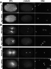
Figure 1.
Figure 1.
The assembly of centrosomal asters in tbg-1(RNAi) embryos correlates with the presence of visibly condensed DNA. Embryos were fixed and stained for MTs (left), γ-tubulin (middle), and DNA (right). All panels are aligned with anterior to the left and posterior to the right. (A) During interphase, before visible DNA condensation, MTs grow from the still unseparated centrosomes in wild-type embryos (top, white arrows). Numerous cytoplasmic MTs are also present. In tbg-1(RNAi) embryos (bottom), cytoplasmic MTs are not affected but no centrosomal asters are detected. Remnants of the spindle from the second meiotic division of the oocyte pronucleus are visible in the anterior of both embryos (black arrows). (B) During mitotic prophase, the centrosomal asters in wild-type embryos have increased in size and are located between the two pronuclei (top, white arrows). In tbg-1(RNAi) embryos, MT asters form around unseparated centrosomes (bottom, arrow) positioned behind the sperm pronucleus. (C) After NEBD, a bipolar spindle is assembled in wild-type embryos (top). In tbg-1(RNAi) embryos, centrosomal asters have increased in size, but a mitotic spindle does not assemble (bottom). The DNA from the oocyte and sperm pronuclei remains in separate masses and does not align on a metaphase plate. Note that in some cases, our γ-tubulin antibody weakly stains condensed chromosomes (C, top middle). This staining does not disappear in tbg-1(RNAi) embryos (B, bottom middle). Bar, 10 μm.
Figure 2.
Figure 2.
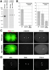
Quantification of γ-tubulin depletion and reduction of centrosomal ZYG-9 in tbg-1(RNAi) embryos. (A) A Western blot of wild-type (left lanes) and tbg-1(RNAi) (right lanes) embryos was probed with antibodies to γ-tubulin (left panel) and α-tubulin (right panel). Bands corresponding to γ- and α-tubulin are indicated (arrowheads). (B) Quantification of centrosomal γ-tubulin and ZYG-9 fluorescence in wild-type (WT) and tbg-1(RNAi) embryos. Centrosomes in eight embryos were quantified for each condition. Total centrosomal fluorescence is expressed in arbitrary units. Error bars are the standard deviation. (C) Sample images from the embryos quantified in B. Wild-type (top) and tbg-1(RNAi) embryos (bottom) were fixed and stained for MTs and DNA (left), γ-tubulin (middle), and ZYG-9 (right). (D) A nocodazole-treated embryo fixed and stained for MTs (left), DNA (middle), and ZYG-9 (right). Bars, 10 μm.
Figure 3.
Figure 3.
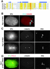
tbg-1(t1465) mutants have a mutation in a residue highly conserved among tubulins. (A) tbg-1(t1465) is a point mutation that leads to the substitution of alanine 401 with valine. A sequence alignment of the COOH termini of α-, β-, and γ-tubulins from S. cerevisiae and C. elegans, α- and β-tubulin from pig, and human γ-tubulin from human is shown. Amino acids conserved in 80–100% of the sequences are highlighted in yellow and alanine 401 is in red. Arrows and tubes above the alignment indicate conserved β sheets and α helices, respectively (Nogales et al., 1998). (B–D) Characterization of the MT cytoskeleton in tbg-1(t1465) embryos. (B) An interphase tbg-1(t1465) embryo stained for MTs (left) and DNA and γ-tubulin (blue and red, right). A remnant of the spindle from the second meiotic division of the oocyte pronucleus is visible (black arrow), but no centrosomal asters are detected. γ-Tubulin localizes to centrosomes (arrows) that fail to associate with the sperm pronucleus (arrowhead). (C) Mitotic tbg-1(t1465) embryos were stained for MTs (left), γ-tubulin (middle), and DNA (right). Two typical examples are shown here (top and bottom). (D) Mitotic tbg-1(t1465+RNAi) embryos stained for MTs and ZYG-9 (green and red, left), γ-tubulin (middle), and DNA (right). Bar, 10 μm.
Figure 4.
Figure 4.
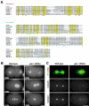
Depletion of CeGrip-1 blocks recruitment of centrosomal γ-tubulin. (A) CeGrip-1 contains the two grip motifs found in proteins that form complexes with γ-tubulin (Gunawardane et al., 2000). By performing further BLAST searches using the grip motifs as a query sequence, we recently identified a second predicted C. elegans protein, CeGrip-2 (C45G3.3), that contains the grip motifs. Here, grip motifs 1 and 2 from CeGrip-1 and CeGrip-2 are aligned with the grip motifs from other proteins homologous to Dgrip91/Spc98p. Amino acids conserved in 80–100% of the sequences are highlighted in yellow. The conserved amino acids that define grip motifs 1 and 2 (Gunawardane et al., 2000) are shown beneath the alignments in red and green, respectively. (B) Panels summarizing a time-lapse sequence of a wild-type (left) and a gip-1(RNAi) embryo (right) expressing both GFP–histone and GFP–γ-tubulin. In gip-1(RNAi) embryos, the γ-tubulin signal was greatly reduced or, as in this case, absent (see Videos 1 and 2 located at http://www.jcb.org/cgi/content/full/jcb.200202047/DC1). (C) Mitotic wild-type and gip-1(RNAi) embryos stained for MTs and DNA (green and red, top), γ-tubulin (middle), and CeGrip (bottom) are shown. CeGrip-1 and γ-tubulin colocalize at centrosomes. Depletion of CeGrip-1 prevents γ-tubulin recruitment to centrosomes and results in an MT phenotype essentially identical to that observed in tbg-1(RNAi) embryos. Depletion of CeGrip-2 also results in an MT phenotype essentially identical to that in tbg-1(RNAi) embryos (unpublished data). Bars, 10 μm.
Figure 5.
Figure 5.
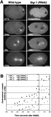
During mitosis in tbg-1(RNAi) embryos, centrosomal α-tubulin fluorescence reaches40% of wild-type levels. (A) Panels summarizing movies of wild-type (left) and tbg-1(RNAi) embryos (right), expressing GFP–α-tubulin. Times are with respect to NEBD. See also Videos 3 and 4 located at http://www.jcb.org/cgi/content/full/jcb.200202047/DC1. In wild type, asters are visible at an early stage (− 477 s). The asters grow in size (all panels) and become the poles of the mitotic spindle (+150 s). (447 s) In tbg-1(RNAi) embryos there are no detectable asters at early time points. (−180 s) Coincident with the onset of mitotic prophase in wild-type (judged by comparison of GFP–α-tubulin and GFP–histone sequences), centrosomal asters form in the tbg-1(RNAi) embryos. At this time, centrosomes (arrow) are often observed to “fly in” to meet the paternal pronucleus. (+189 s) After NEBD, robust mitotic asters form but no spindle assembles. Bar, 10 μm. (B) Kinetic traces of centrosomal α-tubulin fluorescence. Centrosomal fluorescence was quantitated in five wild-type and four tbg-1(RNAi) embryos. Three traces are shown for each.
Figure 6.
Figure 6.
γ-Tubulin is the kinetically dominant centrosomal MT nucleator. Embryos were chilled to depolymerize MTs, rewarmed for the indicated amounts of time, and then fixed and stained to visualize MTs and ZYG-9 (green and red, left in each pair), γ-tubulin (unpublished data), and DNA (right in each pair). Control slides that were not chilled were prepared in parallel (control). After chilling, no MTs are seen emanating from centrosomes in either wild-type or tbg-1(RNAi) embryos (both MT/ZYG-9 and DNA images are projections of entire data stacks). After 15 s regrowth, numerous short MTs emanate from the centrosomes in wild-type embryos, but in tbg-1(RNAi) embryos, only a few centrosomal MTs are observed (these and all subsequent MT/ZYG-9 images are single focal planes). After 30 s regrowth, spindles are found in wild-type embryos (left) and the number and length of MTs have increased in tbg-1(RNAi) embryos (right). After 90 s regrowth, the asters in the tbg-1(RNAi) embryos (right) are similar to asters in unchilled control tbg-1 (RNAi) embryos (bottom right). Bar, 10 μm.
Figure 7.
Figure 7.
Aster formation in γ-tubulin depleted does not require cytoplasmic dynein or ZYG-9. (A) Wild-type, dhc-1(RNAi), and dhc-1(RNAi)+ tbg-1(RNAi) embryos were stained for MTs, DNA, dynein, and γ-tubulin. In wild type (top), dynein (DHC-1) is found in the cytoplasm and enriched around the chromosomes. Depletion of DHC-1 by RNAi (middle) prevents pronuclear migration, centrosome separation, and spindle assembly. Two centrosomal asters form near the posterior cortex of the embryo. Simultaneous depletion of both γ-tubulin and dynein (bottom) results in a qualitatively similar phenotype to dynein depletion alone. (B) Wild-type and tbg-1(RNAi)+zyg-9(RNAi) embryos were stained for MTs, DNA, γ-tubulin, and ZYG-9. In embryos depleted of both γ-tubulin and ZYG-9 (bottom), robust mitotic asters are formed. Bar, 10 μm.

Similar articles

Cited by

References

    1. Alberts, B., D. Bray, J. Lewis, M. Raff, K. Roberts, and J. Watson. 1994. Molecular Biology of the Cell. Garland Publishing, Inc., New York. 1294 pp.
    1. Barbosa, V., R.R. Yamamoto, D.S. Henderson, and D.M. Glover. 2000. Mutation of a Drosophila γ-tubulin ring complex subunit encoded by discs degenerate-4 differentially disrupts centrosomal protein localization. Genes Dev. 14:3126–3139. - PMC - PubMed
    1. Felix, M.A., C. Antony, M. Wright, and B. Maro. 1994. Centrosome assembly in vitro: role of γ-tubulin recruitment in Xenopus sperm aster formation. J. Cell Biol. 124:19–31. - PMC - PubMed
    1. Frankel, F.R. 1976. Organization and energy-dependent growth of microtubules in cells. Proc. Natl. Acad. Sci. USA. 73:2798–2802. - PMC - PubMed
    1. Gaglio, T., M.A. Dionne, and D.A. Compton. 1997. Mitotic spindle poles are organized by structural and motor proteins in addition to centrosomes. J. Cell Biol. 138:1055–1066. - PMC - PubMed

Publication types

MeSH terms