. 2002 May;129(1):122-33.
doi: 10.1104/pp.010986.

Overexpression of polygalacturonase in transgenic apple trees leads to a range of novel phenotypes involving changes in cell adhesion

Affiliations

Overexpression of polygalacturonase in transgenic apple trees leads to a range of novel phenotypes involving changes in cell adhesion

Ross G Atkinson et al. Plant Physiol. 2002 May.

Abstract

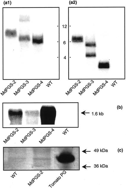
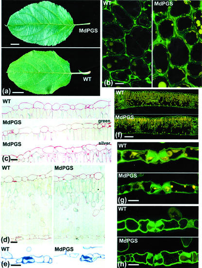
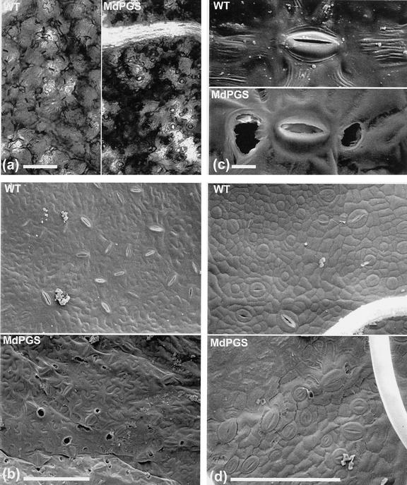
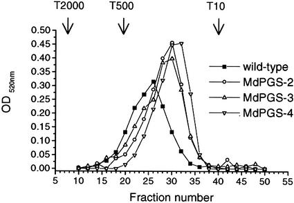
Polygalacturonases (PGs) cleave runs of unesterified GalUA that form homogalacturonan regions along the backbone of pectin. Homogalacturonan-rich pectin is commonly found in the middle lamella region of the wall where two adjacent cells abut and its integrity is important for cell adhesion. Transgenic apple (Malus domestica Borkh. cv Royal Gala) trees were produced that contained additional copies of a fruit-specific apple PG gene under a constitutive promoter. In contrast to previous studies in transgenic tobacco (Nicotiana tabacum) where PG overexpression had no effect on the plant (K.W. Osteryoung, K. Toenjes, B. Hall, V. Winkler, A.B. Bennett [1990] Plant Cell 2: 1239-1248), PG overexpression in transgenic apple led to a range of novel phenotypes. These phenotypes included silvery colored leaves and premature leaf shedding due to reduced cell adhesion in leaf abscission zones. Mature leaves had malformed and malfunctioning stomata that perturbed water relations and contributed to a brittle leaf phenotype. Chemical and ultrastructural analyses were used to relate the phenotypic changes to pectin changes in the leaf cell walls. The modification of apple trees by a single PG gene has offered a new and unexpected perspective on the role of pectin and cell wall adhesion in leaf morphology and stomatal development.


Figures

Figure 1
Figure 1
Analysis of MdPGS transformant and wild-type (WT) apple plants by DNA gel blot, RNA gel blot, and western blot. a, DNA gel-blot analysis was performed using genomic DNA (10 μg) digested with BamHI and size fractionated on 0.8% (w/v) agarose gels. Membranes were probed at the left border with an NptII-specific probe (a1) and at the right border with a cauliflower mosaic virus 35S-specific probe (a2). Membranes were washed at a stringency allowing 15% mismatch (0.5× SSC and 1% [w/v] SDS, 65°C). Size markers are in kb. b, RNA gel-blot analysis was performed using RNA (5 μg) probed with a GDPG1-specific probe (GDPG1 nucleotides 1–603). Membranes were washed at a final stringency of 0.1× SSC + 0.1% (w/v) SDS and exposed for 16 h. Signals were analyzed using a phospho-imaging system and ImageQuant software analysis and corrected for any small differences in RNA loading by comparison with ribosomal RNA hybridization. c, For western-blot analysis, PG enzyme was extracted as described in “Materials and Methods” and separated on denaturing 10% (w/v) polyacrylamide gels (loading approximately 11 μg protein per lane). Blots were incubated with antibodies raised against endo-PG from ripening tomato fruit to detect cross-reacting proteins. Endo-PG purified from ripening tomato fruit (DellaPenna et al., 1986) was used as a positive control. Arrows indicate position of Mr markers.
Figure 2
Figure 2
Phenotype of leaves from MDPGS transformants compared with wild type (WT). a, Silvery leaf phenotype. Scale bar = 10 mm. b, Putative petiole abscission zone showing distribution of low-esterified pectin by labeling with JIM5 antibodies. c, Cross section of upper epidermis and palisade cells of leaves demonstrating reduced cell adhesion in the silvery leaf phenotype (stained with ruthenium red for pectin). d, Cross section of entire leaves, demonstrating the reduced ruthenium red staining of the lower epidermis and holes next to guard cells or stomata. e, Cross section of the lower leaf epidermis showing the hole next to the guard cell in the transformant (stained with toluidine blue). f, Cross section of leaves labeled with JIM5 antibodies, showing reduced labeling of lower epidermal cells in the transformant. g, Cross section of the lower epidermis of leaves labeled with JIM5 antibodies showing altered distribution of label in guard cells of transformant stomata. h, Cross section of the lower epidermis of leaves labeled with JIM7 antibodies showing labeling in the guard cells and neighboring cells. Scale bars in b through h = 10 μm.
Figure 3
Figure 3
Stomata from MdPGS transformant leaves compared with wild type (WT). a, Light microscope view of stomata 30 min after placement in the dark. b and c, Holes associated with stomata in mature transgenic leaves compared with wild-type. d, Stomata in young, developing leaves of transgenic and wild-type plants. Scale bars in a, b, and d = 100 μm; scale bar in c = 10 μm.
Figure 4
Figure 4
Molecular distribution of CDTA-soluble pectin. Mr distribution was analyzed by gel-permeation chromatography on a Sepharose CL-2B column (65 × 2 cm; eluent 0.05 m Na-acetate, pH 6.0; 0.125 m NaCl; 0.05% [w/v] chlorobutanol; fraction size 20 min; flow rate 8.4 mL h−1). The column was calibrated using dextran molecular standards T2000, T500, and T10 (Pharmacia Biotech, Piscataway, NJ), and the elution profile monitored using the GalUA assay (Ahmed and Labavitch, 1977). Na2CO3-soluble pectins of all genotypes were not soluble after freeze drying and therefore could not be subjected to gel permeation chromatography.

Similar articles

Cited by

References

    1. Ahmed AER, Labavitch JM. A simplified method for accurate determination of cell wall uronide content. J Food Biochem. 1977;1:361–365.
    1. Albersheim P, Nevins DJ, English PD, Karr A. A method for the analysis of sugars in plant cell-wall polysaccharides by gas-liquid chromatography. Carbohydr Res. 1967;5:340–345.
    1. ASTM. ASTM Standards on Color and Appearance Measurement. Ed 5. West Conshohocken, PA: ASTM; 1996.
    1. Atkinson RG. A cDNA clone for endopolygalacturonase from apple. Plant Physiol. 1994;105:1437–1438. - PMC - PubMed
    1. Atkinson RG, Bolitho KM, Wright MA, Iturriagagoitia-Bueno T, Reid SJ, Ross GS. Apple ACC-oxidase and polygalacturonase: ripening-specific gene expression and promoter analysis in transgenic tomato. Plant Mol Biol. 1998;38:449–460. - PubMed

Publication types

MeSH terms

LinkOut - more resources