. 2002 May 14;99(10):6673-8.
doi: 10.1073/pnas.102167799.

Preferential binding of ATR protein to UV-damaged DNA

Affiliations

Preferential binding of ATR protein to UV-damaged DNA

Keziban Unsal-Kaçmaz et al. Proc Natl Acad Sci U S A. .

Abstract

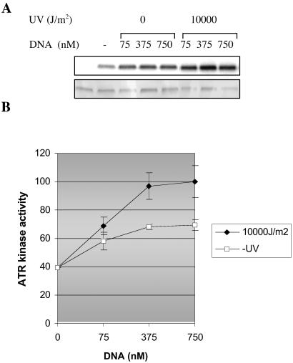
The ATR protein is a member of the phosphoinositide 3-kinase-related kinase family and plays an important role in UV-induced DNA damage checkpoint response. Its role as a signal transducer in cell cycle checkpoint is well established, but it is currently unclear whether ATR functions as a damage sensor as well. Here we have purified the ATR protein and investigated its interaction with DNA by using biochemical analysis and electron microscopy. We find that ATR is a DNA-binding protein with higher affinity to UV-damaged than undamaged DNA. In addition, damaged DNA stimulates the kinase activity of ATR to a significantly higher level than undamaged DNA. Our data suggest that ATR may function as an initial sensor in the DNA damage checkpoint response.


Figures

Figure 1
Figure 1
Purification of full-length and various fragments of ATR from transiently transfected HEK293T cells. (A) Schematic representation of the full-length ATR (ATR-F) and its N-terminal (ATR-N), middle region (ATR-M), and C-terminal (ATR-C) fragments. (B) Recombinant wild-type (wt) and kinase-dead (kd) ATR, each containing a Flag affinity tag, purified from HEK293T cells, were separated by SDS/PAGE and visualized by silver staining shown (Left). Western blot of the ATR preparations with an antibody specific for the N-terminal FLAG tag is shown (Right). For the kinase activity of recombinant wild-type and kinase-dead ATR, purified proteins were incubated with [γ-32P]-ATP, and reaction products were electrophoresed and visualized by silver staining (Bottom) or autoradiography (Top). (C) Expression of Flag-epitope tagged and affinity-purified ATR-fragments as analyzed by silver staining.
Figure 2
Figure 2
ATR binds DNA. HEK293T cells were transfected with vector expressing full-length ATR (ATR-F), its N-terminal (ATR-N), middle region (ATR-M), and C-terminal (ATR-C) fragments or the nontransfected (Mock) cells were lysed and the tagged proteins were purified by binding to FLAG-agarose beads. The beads were then incubated with radiolabeled DNA substrate as described in Materials and Methods. After exhaustive washing, the beads were loaded onto a 10% SDS/PAGE, and the bound proteins were visualized by silver staining (Top), and the levels of ATR-bound substrates were visualized by autoradiography (Middle). Bottom shows the quantitative analysis of data from Middle.
Figure 3
Figure 3
UV crosslinking of full-length ATR with DNA. Purified full-length ATR (ATR-F) and its fragments, ATR-N, ATR-M, and ATR-C, were mixed with the azidophenacetyl-derivatized DNA probe, and the reaction mixtures were exposed to 366-nm light on ice. The reaction products were separated by SDS/PAGE and analyzed by (A) silver staining and (B) autoradiography.
Figure 4
Figure 4
ATR preferentially binds to UV (–4) photoproduct. HEK293T cells transfected with a vector expressing full-length ATR or no transfected (Mock) cells were lysed and the tagged proteins were purified by binding to FLAG-agarose beads. Proteins bound to beads were incubated with either unmodified (U) 46-mer duplex or (–4)-photoproduct containing duplex of the same sequence (M). After extensive washing, the material bound to the beads was analyzed by SDS/PAGE followed by silver staining (Top) and autoradiography (Middle). The input lanes contain 1/10th of the DNA used in the binding assay. Bottom shows the mean values and standard errors of DNA binding calculated from three independent experiments.
Figure 5
Figure 5
ATR-DNA binding analyzed by electron microscopy. (A) Visualization of the binding of ATR protein to (a) linearized or (b) supercoiled pGEMEX1 dsDNA. The samples were directly mounted onto thin carbon-coated foils and rotary shadowed with tungsten. The complexes shown in a and b are representatives of the total population of protein-bound DNA molecules observed. (Bar = 250 nm.) (B) Quantitative comparison of ATR DNA binding to unirradiated or UV-irradiated plasmid DNA. The number of protein–DNA complex is given in relation to the number of DNA molecules counted in each reaction.
Figure 6
Figure 6
Phosphorylation of p53 by purified ATR is stimulated by damaged DNA. (A) Kinase reactions were performed with p53 alone (lane 1) or p53 and ATR in the absence of DNA (lane 2), in the presence of untreated DNA (lanes 3–5), or in the presence of UV-treated DNA (lanes 6–8), as indicated. Proteins were separated by SDS/PAGE and visualized by silver staining (Bottom), and phosphorylated p53 was detected by PhosphorImager (Top). (B) Quantitative analysis of ATR kinase activity. The mean values and standard errors were calculated from three independent experiments. The relative kinase activity of ATR is normalized against the highest kinase activity obtained, which was at UV irradiated DNA concentration of 750 nM.

Similar articles

Cited by

References

    1. Abraham R T. Genes Dev. 2001;15:2177–2196. - PubMed
    1. Lowndes N F, Murguia J R. Curr Opin Genet Dev. 2000;10:17–25. - PubMed
    1. Zhou B B, Elledge S J. Nature (London) 2000;408:433–439. - PubMed
    1. Shiloh Y. Curr Opin Genet Dev. 2001;11:71–77. - PubMed
    1. Durocher D, Jackson S P. Curr Opin Cell Biol. 2001;13:225–231. - PubMed

Publication types

MeSH terms