. 2002 May 14;99(10):6889-94.
doi: 10.1073/pnas.072190799.

ARGONAUTE1 is required for efficient RNA interference in Drosophila embryos

Affiliations

ARGONAUTE1 is required for efficient RNA interference in Drosophila embryos

Robert W Williams et al. Proc Natl Acad Sci U S A. .

Abstract

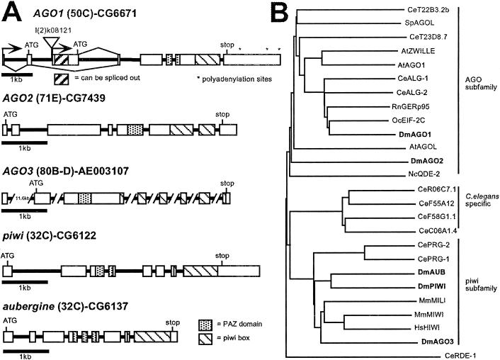
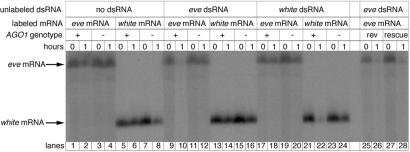
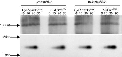
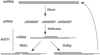
Double-stranded RNA (dsRNA) triggers homology-dependent posttranscriptional gene interference (RNAi) in a diverse range of eukaryotic organisms, in a process mechanistically related to viral and transgene-mediated cosuppression. RNAi is characterized by the conversion of long dsRNA into approximately 21-25-nt small interfering RNAs (siRNA) that guide the degradation of homologous mRNA. Many of the genes required for siRNA production and target mRNA degradation are widely conserved. Notably, members of the Argonaute-like gene family from Arabidopsis, Caenorhabditis elegans, Drosophila, and Neurospora have been genetically and/or biochemically identified as components of the RNAi/cosuppression pathway. We show here that mutations in the Drosophila Argonaute1 (AGO1) gene suppress RNAi in embryos. This defect corresponds to a reduced ability to degrade mRNA in response to dsRNA in vitro. Furthermore, AGO1 is not required for siRNA production in vitro nor can the introduction of siRNA bypass AGO1 mutants in vivo. These data suggest that AGO1 functions downstream of siRNA production.


Figures

Figure 1
Figure 1
The Argonaute gene family. (A) Genomic organization and cytological location of the five Drosophila Argonaute-like genes. Exons are shown as boxes and introns are shown as solid lines. The PAZ and PIWI domains are defined by Cerutti et al. (31). The AGO1 gene has two transcriptional start sites, resulting in two different ORFs differing in the first 65 amino acids. The AGO1 P element insertion l(2)k08121 is shown. l(2)k16601, an independent insertion in AGO1, is located 8 base pairs downstream from the l(2)k08121 insertion site. AGO3 is located near the centromere at 80B-D. Only one of the seven introns has been completely sequenced. The curated gene name, CG, is given for all except AGO3. The GenBank accession no. for a genomic scaffold sequence that contains AGO3 is listed instead. (B) Phylogenetic grouping of the Drosophila Argonaute-like proteins and representatives from other organisms. The tree was constructed with the full-length protein sequences with CLUSTALX and a BLOSUM protein weight matrix. Dm, Drosophila melanogaster; Ce, C. elegans; At, Arabidopsis thaliana; Sp, Schizosaccharomyces pombe; Mm, Mus musculus; Hs, Homo sapiens; Oc, Oryctolagus cuniculus; Rn, Rattus norvegicus. The Drosophila sequences are shown in bold.
Figure 2
Figure 2
Embryonic expression patterns of the five Drosophila Argonaute-like genes. (AD) AGO1, (EH) AGO2, (IL) AGO3, (MP) piwi, and (QT) aub. (A, E, I, M, and Q) Stage 4–5 embryos. (B and F) Stage 6 embryos initiating gastrulation. (C and G) Stage 8 embryos. (D and H) Stage 15–16. (J, N, and R) Stage 10–12 embryos. (K, O, and S) Stage 14. (L, P, and T) Stage 15–16. Anterior is to the left. Lateral views in A, C, E, G, I, J, M, N, Q, and R. Ventral view in B, D, F, and H. Dorsal view in K, L, O, P, S, and T.
Figure 3
Figure 3
AGO1 mutant embryos have a reduced response to eve dsRNA. Representative embryonic phenotypes of wild-type, GFP-expressing AGO1+, and AGO1k08121 mutant embryos injected with dsRNA are shown. (A) w1118 embryo injected with white dsRNA showing no alteration in the number of ventral denticle belts. (B) w1118 embryo injected with eve dsRNA. Denticle belt 4 is fused with 5, and 6 is missing. (C) AGO1+ embryo injected with eve dsRNA. Belt 6 is missing. (D) AGO1k08121 embryo injected with eve dsRNA showing no alteration in the number of denticle belts. (E) AGO1+ embryo injected with eve siRNA. Belt 4 is partially formed whereas belt 6 is missing. (F) AGO1k08121 embryo injected with eve siRNA showing no alteration in the number of denticle belts. The denticle belts corresponding to each abdominal segment are labeled, except 8, which is not in the plane of focus.
Figure 4
Figure 4
Degradation of targeted mRNA in response to dsRNA is reduced in AGO1 mutants. Gel of uniformly 32P-labeled mRNA that was incubated in extracts prepared from AGO1k08121 (−) and GFP-expressing AGO1+ (+) embryos. Before the addition of labeled mRNA, the extracts were incubated with or without unlabeled dsRNA. Samples collected just after the addition of mRNA (0) or after a 1-h incubation (1) are in neighboring lanes. There is some nonspecific degradation of the mRNA after 1 h in the embryo extracts as seen in the “no dsRNA” controls. However, the amount of mRNA is greatly reduced in (+) extracts preincubated with homologous dsRNA. mRNA is unaffected in extracts prepared from AGO1 mutants. Extracts prepared from a viable revertant line derived from l(2)k08121 (rev) are able to degrade targeted mRNA. Similarly, extracts prepared from AGO1k08121 embryos rescued with an AGO1 cDNA (rescue) degrade mRNA targeted with homologous dsRNA.
Figure 5
Figure 5
Production of ≈21-nt fragments is unaffected in AGO1 mutants. Gel of uniformly 32P-labeled dsRNA, corresponding to either the eve or white gene, which was incubated in extracts from GFP-expressing AGO1+ embryos and AGO1k08121 mutant embryos for 0, 10, 20, and 30 min. Upper is the top part of the gel showing the full-length input RNA. Lower shows the lower part of the same gel showing that both eve dsRNA and white dsRNA were cleaved into ≈21-nt fragments.
Figure 6
Figure 6
Proposed step(s) during RNAi where AGO1 may function. RNAi is initiated by the Dicer-mediated cleavage of dsRNA into ≈21-nt siRNA. siRNA is then used to target mRNA for degradation by the RISC complex. In another step, which may or may not be mediated by RISC, siRNA can act as a primer for second-strand synthesis of RNA complementary to the targeted mRNA. The newly dsRNA may then become a substrate for Dicer. Based on our results, we proposed that AGO1 functions downstream of siRNA production. Our experiments do not address whether AGO1 is specific for mRNA degradation or whether it is required for the RNA-dependent RNA polymerase (RdRp) amplification of the siRNA.

References

    1. Hammond S M, Caudy A A, Hannon G J. Nat Rev Genet. 2001;2:110–119. - PubMed
    1. Cogoni C, Macino G. Proc Natl Acad Sci USA. 1997;94:10233–10238. - PMC - PubMed
    1. Fire A, Xu S, Montgomery M K, Kostas S A, Driver S E, Mello C C. Nature (London) 1998;391:806–811. - PubMed
    1. Grishok A, Pasquinelli A E, Conte D, Li N, Parrish S, Ha I, Baillie D L, Fire A, Ruvkun G, Mello C C. Cell. 2001;106:23–34. - PubMed
    1. Hutvagner G, McLachlan J, Pasquinelli A E, Balint E, Tuschl T, Zamore P D. Science. 2001;293:834–838. - PubMed

Publication types