Postnatal refinement of auditory nerve projections to the cochlear nucleus in cats
- PMID: 12012373
- PMCID: PMC2386504
- DOI: 10.1002/cne.10176
Postnatal refinement of auditory nerve projections to the cochlear nucleus in cats
Abstract
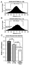
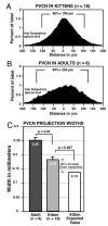
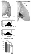
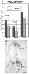
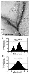
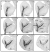
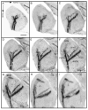
Studies of visual system development have suggested that competition driven by activity is essential for refinement of initial topographically diffuse neuronal projections into their precise adult patterns. This has led to the assertion that this process may shape development of topographic connections throughout the nervous system. Because the cat auditory system is very immature at birth, with auditory nerve neurons initially exhibiting very low or no spontaneous activity, we hypothesized that the auditory nerve fibers might initially form topographically broad projections within the cochlear nuclei (CN), which later would become topographically precise at the time when adult-like frequency selectivity develops. In this study, we made restricted injections of Neurobiotin, which labeled small sectors (300-500 microm) of the cochlear spiral ganglion, to study the projections of auditory nerve fibers representing a narrow band of frequencies. Results showed that projections from the basal cochlea to the CN are tonotopically organized in neonates, many days before the onset of functional hearing and even prior to the development of spontaneous activity in the auditory nerve. However, results also demonstrated that significant refinement of the topographic specificity of the primary afferent axons of the auditory nerve occurs in late gestation or early postnatal development. Projections to all three subdivisions of the CN exhibit clear tonotopic organization at or before birth, but the topographic restriction of fibers into frequency band laminae is significantly less precise in perinatal kittens than in adult cats. Two injections spaced > or = 2 mm apart in the cochlea resulted in labeled bands of projecting axons in the anteroventral CN that were 53% broader than would be expected if they were proportional to those in adults, and the two projections were incompletely segregated in the youngest animals studied. Posteroventral CN (PVCN) projections (normalized for CN size) were 36% broader in neonates than in adults, and projections from double injections in the youngest subjects were nearly fused in the PVCN. Projections to the dorsal division of the CN were 32% broader in neonates than in adults when normalized, but the dorsal CN projections were always discrete, even at the earliest ages studied.
Copyright 2002 Wiley-Liss, Inc.
Figures



Comment in
-
Choosing axonal real estate: location, location, location.J Comp Neurol. 2002 Jun 17;448(1):1-5. doi: 10.1002/cne.10255. J Comp Neurol. 2002. PMID: 12012372 Review. No abstract available.
References
-
- Altman J, Bayer SA. Development of the brain stem in the rat. III. Thymidine-radiographic study of the time of origin of neurons of the vestibular and auditory nuclei of the upper medulla. J. Comp. Neurol. 1980;194:877–904. - PubMed
-
- Bourk TR, Mielcarz JM, Norris BE. Tonotopic organization of the anteroventral cochlear nucleus of the cat. Hearing Res. 1981;4:215–241. - PubMed
-
- Brawer JR, Morest DK, Kane EI. The neuronal architecture of the cochlear nucleus of the cat. J. Comp. Neurol. 1974;155:251–300. - PubMed
-
- Brugge JF, Reale RA, Wilson GF. Sensitivity of auditory cortical neurons of kittens to monaural and binaural high frequency sound. Hearing Res. 1988;34:127–140. - PubMed
-
- Cant NB, Morest DK. The structural basis for stimulus coding in the cochlear nucleus of the cat. In: Berlin C, editor. Hearing Science: Recent Advances. College-Hill; San Diego: 1984. pp. 371–421.
Publication types
MeSH terms
Substances
Grants and funding
LinkOut - more resources
Full Text Sources
Miscellaneous

