. 2002 Jun;129(11):2619-28.
doi: 10.1242/dev.129.11.2619.

The zinc-finger transcription factor Klf4 is required for terminal differentiation of goblet cells in the colon

Affiliations

The zinc-finger transcription factor Klf4 is required for terminal differentiation of goblet cells in the colon

Jonathan P Katz et al. Development. 2002 Jun.

Abstract

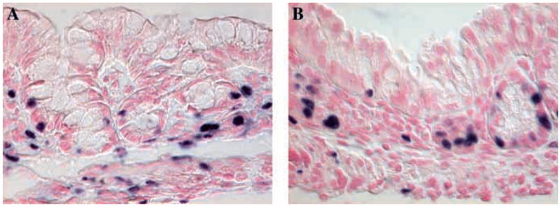
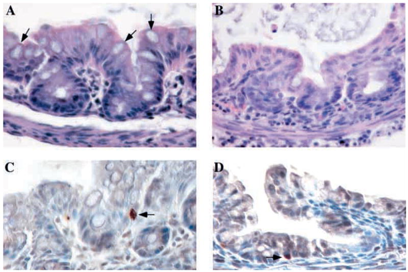
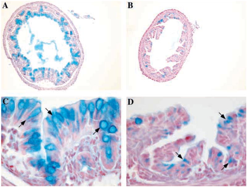
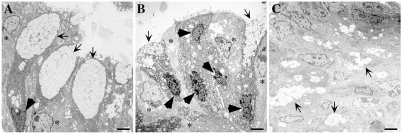
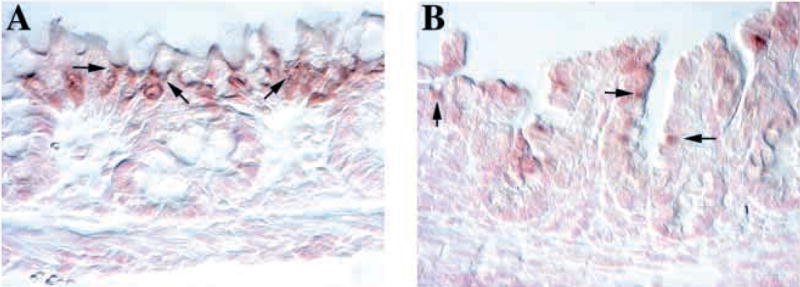
Klf4 (formerly GKLF) is a zinc-finger transcription factor expressed in the epithelia of the skin, lungs, gastrointestinal tract and several other organs. In vitro studies have suggested that Klf4 plays an important role in cell proliferation and/or differentiation. Mice homozygous for a null mutation in Klf4 die within 15 hours of birth and show selective perturbation of late-stage differentiation structures in the epidermis, but the function of Klf4 in the gastrointestinal tract has not been investigated. To address this issue, we have generated Klf4(-/-) mice by homologous recombination in embryonic stem cells. In this study, we provide the first in vivo evidence that Klf4 is a goblet cell-specific differentiation factor in the colon. Klf4(-/-) mice exhibit normal cell proliferation and cell death rates in the colon on postnatal day 1. However, Klf4(-/-) mice demonstrate a 90% decrease in the number of goblet cells in the colon, show abnormal expression of the goblet cell-specific marker Muc2 by in situ hybridization, have abnormal staining of the colonic epithelium with Alcian Blue for acidic mucins, and lack normal goblet cell morphology by ultrastructural analysis. All other epithelial cell types are present in the colon of Klf4(-/-) mice. In summary, Klf4 plays a crucial role in colonic epithelial cell differentiation in vivo.


Figures

Fig. 1
Fig. 1
Targeting strategy and in situ hybridization for Klf4. (A) The four alleles used in the gene targeting of Klf4 are shown with predicted band sizes of restriction fragments used in Southern blot analysis. Deletion of exons 2 and 3 produces a frame shift in exon 4. Exon 1 encodes only the first amino acid of the Klf4 protein. A 400 bp fragment external to the targeting vector was used as the probe for Southern blot. White arrows depict PCR primer locations. (B) Southern blot showing the wild-type and targeted Klf4 bands. (C) PCR screening for Klf4 revealed 172 bp wild-type, 296 bp floxed and 425 bp null bands. (D,E) In situ hybridization for Klf4 in colon from postnatal day 1 mice. (D) Wild-type mice demonstrated expression of Klf4 mRNA (arrows) throughout the epithelium. (E) Klf4−/− mice showed no expression of Klf4 mRNA, confirming the successful deletion of Klf4. E, EcoRI; X, XbaI.
Fig. 2
Fig. 2
(A,B) Immunohistochemistry for BrdU, indicating dividing cells in the colonic epithelium of postnatal day 1 mice. Klf4−/−mice (B) were no different from wild-type (A) littermates in rates of cell proliferation in the colonic epithelium.
Fig. 3
Fig. 3. Differentiation of goblet cells but not colonocytes and enteroendocrine cells was abnormal in Klf4−/− mice. (A,B) Hematoxylin and Eosin staining of the colon from postnatal day 1 mice
(A) The colonic epithelium of wild-type mice showed a normal contour and numerous goblet cells (arrows) along the crypts and surface epithelium. (B) By contrast, goblet cells were nearly absent from Klf4−/− epithelium, and mutant mice appeared to have subtle changes in epithelial contour. Colonocytes appeared grossly normal. (C,D) Immunohistochemistry for chromogranin A, a marker of enteroendocrine cells, in colon from postnatal day 1 mice. Enteroendocrine cells (arrows) were identified in both wild-type (C) and Klf4−/−mice (D). In both cases, enteroendocrine cells were seen at typically low numbers, with only one to two enteroendocrine cells per transverse section.
Fig. 4
Fig. 4
Klf4−/− mice showed a dramatic decrease in the number of goblet cells in the colon. (A–D) Alcian Blue staining for acidic mucins in the colon of postnatal day 1 mice. At low power, wild-type mice (A) had numerous Alcian Blue-positive cells, but very few Alcian Blue positive cells were seen in the Klf4−/−mice (B). At higher power, Alcian Blue staining was seen only in the goblet cells (arrows) of wild-type mice (C), while Klf4−/− mice (D) displayed numerous epithelial cells with small amounts of Alcian Blue staining material (arrows) but no normal goblet cell morphology.
Fig. 5
Fig. 5
Electron microscopy revealed abnormal ultrastructural morphology in the colonic epithelium of Klf4−/− mice. (A) Colonic epithelium from wild-type mice on postnatal day 1 showed the normal goblet cell morphology (arrows). The nucleus (black arrowhead) was densely packed and located at the base of the cell, while the apical portion of the cell was comprised largely of membrane-bound secretory vacuoles containing mucigen. Normal colonocytes (*) were interspersed. (B,C) Colonic epithelium from Klf4−/− mice on postnatal day 1. (B) Several abnormally shaped or immature cells of the goblet lineage (arrows) were seen emptying their contents into the lumen. Adjacent to these cells, several cells had densely packed nuclei (black arrowheads) suggestive of a goblet cell lineage but lacked the typical secretory vacuoles and normal goblet cell shape. Colonocytes (*), recognized by their paler cytoplasm and less condensed nuclei, appeared normal. (C) The colonic epithelium of the Klf4−/− mouse also demonstrated small clusters of secretory vacuoles (arrows) scattered throughout the epithelium. Scale bars: 2 μm.
Fig. 6
Fig. 6
Altered expression of the goblet cell marker Muc2 in Klf4−/− mice. (A,B) In situ hybridization for Muc2 in the colon at postnatal day 1. (A) Wild-type mice showed a goblet-cell specific expression pattern of Muc2 mRNA (arrows) with abundant expression adjacent to the apical mucigen granules. Staining was prominent only in surface epithelial cells. (B) Klf4−/− mice had expression of Muc2 mRNA (arrows) in the epithelium, but this expression was not confined to cells with goblet morphology. Patchy expression was seen in cells throughout the epithelium.

References

    1. Bach SP, Renehan AG, Potten CS. Stem cells: the intestinal stem cell as a paradigm. Carcinogenesis. 2000;21:469–476. - PubMed
    1. Bieker JJ. Krüppel-like factors: three fingers in many pies. J Biol Chem. 2001;276:34355–34358. - PubMed
    1. Burkitt HG, Young B, Heath JW. Wheater’s Functional Histology: A Text and Color Atlas. New York: Churchill Livingstone; 1993.
    1. Chang WW, Leblond CP. Renewal of the epithelium in the descending colon of the mouse. I Presence of three cell populations: vacuolated-columnar, mucous and argentaffin. Am J Anat. 1971;131:73–99. - PubMed
    1. Chang WW, Nadler NJ. Renewal of the epithelium in the descending colon of the mouse. IV Cell population kinetics of vacuolated-columnar and mucous cells. Am J Anat. 1975;144:39–56. - PubMed

Publication types

MeSH terms