. 2002 May 15;541(Pt 1):319-31.
doi: 10.1113/jphysiol.2002.016857.

Cortical sensory suppression during arousal is due to the activity-dependent depression of thalamocortical synapses

Affiliations

Cortical sensory suppression during arousal is due to the activity-dependent depression of thalamocortical synapses

Manuel A Castro-Alamancos et al. J Physiol. .

Abstract

The thalamus serves as a gate that regulates the flow of sensory inputs to the neocortex, and this gate is controlled by neuromodulators from the brainstem reticular formation that are released during arousal. Here we show in rats that sensory-evoked responses were suppressed in the neocortex by activating the brainstem reticular formation and during natural arousal. Sensory suppression occurred at the thalamocortical connection and was a consequence of the activity-dependent depression of thalamocortical synapses caused by increased thalamocortical tonic firing during arousal. Thalamocortical suppression may serve as a mechanism to focus sensory inputs to their appropriate representations in neocortex, which is helpful for the spatial processing of sensory information.


Figures
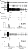
Figure 1
Figure 1. Activation induced by RF stimulation produces sensory suppression in neocortex
A, field potential (FP) and single-unit recordings obtained in the barrel cortex through the same electrode, and a simultaneously recorded single unit in the VPM thalamus of a urethane-anaesthetized rat. RF stimulation was delivered for 1 s (100 Hz) and produced a robust activating effect consisting of low amplitude irregular activity in the cortical field potential, reduced firing in the cortical unit and enhanced firing in the VPM unit. B, raw traces and binned sum data from 14 trials of sensory responses evoked by whisker stimulation before (Control) and after RF stimulation. The cortical field and unit responses are suppressed by RF stimulation, while the thalamic unit response is enhanced. C, cortical single-unit recording obtained in the same experiment shown in A and B. In contrast to cortical unit 1 shown in A and B, cortical unit 2 responds to RF stimulation by increasing its firing rate. However, like cortical unit 1 this unit also suppresses its response to whisker stimulation. Cortical unit 2 was recorded after cortical unit 1 in the same penetration; the thalamic unit was the same for both cases.
Figure 3
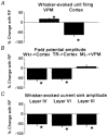
Figure 3. Population data showing the percentage changes induced by RF stimulation of VPM and cortex responses
A, percentage changes induced by RF stimulation of VPM and cortex single-unit firing probability to whisker stimulation at short latency intervals (3-7 ms for VPM and 5-12 ms for cortex). n = 55 and 65 units per group, respectively. *P < 0.0001, t test. B, percentage changes induced by RF stimulation of field potential responses evoked in cortex by whisker stimulation (Wkr → Cortex) or thalamic radiation stimulation (TR → Cortex) and of responses evoked in VPM by medial lemniscus stimulation (ML → VPM). n = 15, 6 and 5 experiments per group, respectively. *P < 0.0001, t test. C, percentage changes induced by RF stimulation of current sink amplitudes evoked by whisker stimulation in layer IV, layer VI and layer III. n = 3 experiments per group. *P < 0.0001, t test.
Figure 2
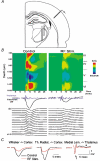
Figure 2. Sensory suppression during activation occurs at the thalamocortical connection
A, schematic representation of the location of the 16-channel silicon probe placed at a 45 deg angle in the barrel cortex, which was used to record field potential responses through the layers of barrel neocortex. Also note a single recording electrode placed in the VPM thalamus and a microdialysis probe located adjacent to the recording electrode. The microdialysis probe was used to infuse TTX into the VPM as described in Fig. 5. B, current source-density analysis (CSD) of the sensory response evoked in the barrel cortex by whisker stimulation before (Control) and after RF stimulation. The sink (red) and source (blue) distribution reveals that the short latency responses in layers VI and IV are strongly depressed by RF stimulation. Also shown below is multiunit activity from the VPM thalamus and a field potential recording from one of the cortical sites (900 μm in depth). The multiunit traces are the average of five sensory responses. Notice the depression of the cortical response, but not of the thalamic response, after RF stimulation. The field potentials used to derive the CSD are shown at the bottom. The scale range for the CSD is +3.5 to −3.5 mV mm−2. C, overlaid field potential responses showing the effect of RF stimulation (red traces) on cortical responses evoked by whisker stimulation (left), cortical responses evoked by thalamic radiation stimulation (middle) and on VPM responses evoked by medial lemniscus stimulation (right). The lemniscal response has two components, marked by an arrow and an asterisk (see text for details). The responses are the average of ten traces.
Figure 5
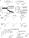
Figure 5. Sensory suppression induced by RF stimulation is abolished by thalamic inactivation
A, cortical field potential responses to whisker stimulation (left traces) and to stimulation of the thalamic radiation (right traces). The arrows mark the onset of the whisker stimulus (left) and the thalamic radiation electrical stimulus (right). The numbers on the traces mark the locations on the plot below. Infusion of TTX into the VPM thalamus abolishes the cortical response to whisker stimulation, but not the cortical response to thalamic radiation stimulation. Also shown (right) is a power-spectrum of the field potential activity recorded in the cortex before (Control) and after RF stimulation (RF stim) when the thalamus was intact (continuous line) or inactivated with TTX (dashed line). Thalamic inactivation does not significantly affect the cortical activating effect of RF stimulation. B, field potential responses to thalamic radiation stimulation are suppressed by RF stimulation when the thalamus is intact, but not when it is inactivated with TTX. C, the thalamocortical response evoked by stimulating the thalamic radiation is suppressed by activity. Repetitive stimulation of the thalamic radiation at 10 Hz sharply depresses the thalamocortical response (left), and this effect is equivalent to RF stimulation in an intact thalamus (right). The asterisk marks the small and long latency response presumed to be due to intracortical collaterals of corticothalamic cells (see Discussion for details).
Figure 4
Figure 4. Natural arousal produces thalamocortical suppression
A, fast Fourier transform (FFT) of the spontaneous field potential activity recorded from the barrel cortex of a freely behaving rat. Blue indicates low power and red indicates high power for the frequency on the y-axis. B, top: amplitude of the thalamocortical response evoked in the barrel cortex by stimulating the thalamic radiation every 10 s (open circles). The running averages of three successive responses are shown by filled circles. Middle, amplitude of the electromyographic activity (EMG; arbitrary units) recorded from the whisker pad with subcutaneous electrodes. Bottom, locomotor activity (arbitrary units) recorded by photobeam detectors in the cage. The x-axis time scale corresponds to all graphs. The animal is sleeping for the initial 11 min (i.e. lying down in the cage with eyes closed) and the amplitude of the thalamocortical response is large. After 11 min, the rat wakes up and moves actively about the cage for the remainder of the experiment, and the thalamocortical response is suppressed. C, traces correspond to a thalamocortical response evoked during slow-wave sleep and during the active exploratory state that follows. Each trace shown is 32.5 ms. The arrows mark the onset of the electrical stimulus to the thalamic radiation.
Figure 6
Figure 6. Blocking cholinergic, noradrenergic and GABAB receptors in the neocortex does not abolish sensory suppression induced by RF stimulation
A, field potential responses evoked in the neocortex by whisker stimulation. Under control conditions RF stimulation suppresses the evoked response (upper traces). Simultaneous application of scopolamine, hexamethodide, phentolamine, propanolol and CGP35348 enhances the whisker-evoked response, but under these conditions RF stimulation also suppresses the sensory-evoked response (lower traces). Traces are the average of five responses from a representative experiment. B, population data from three experiments in which the drugs mentioned in A were applied. The average for each experiment was calculated from 10-15 control traces and RF traces. RF significantly suppresses whisker-evoked responses during control conditions and after application of the drug combination (*P < 0.0001, t test). Also, application of the drugs significantly enhances the evoked response as compared to control (**P < 0.0001, t test).

References

    1. Abbott LF, Varela JA, Sen K, Nelson SB. Synaptic depression and cortical gain control. Science. 1997;275:220–224. - PubMed
    1. Armstrong-James M. The nature and plasticity of sensory processing within adult rat barrel cortex. In: Jones EG, Diamond IT, editors. Cerebral Cortex, The Barrel Cortex of Rodents. Vol. 11. New York: Plenum; 1995. pp. 333–374.
    1. Armstrong-James M, Fox K. Spatiotemporal convergence and divergence in the rat S1 ‘barrel’ cortex. Journal of Comparative Neurology. 1987;263:265–281. - PubMed
    1. Armstrong-James M, Fox K, Das-Gupta A. Flow of excitation within rat barrel cortex on striking a single vibrissa. Journal of Neurophysiology. 1992;68:1345–1358. - PubMed
    1. Aston-Jones G, Chiang C, Alexinsky T. Discharge of noradrenergic locus coeruleus neurons in behaving rats and monkeys suggests a role in vigilance. Progress in Brain Research. 1991;88:501–520. - PubMed

Publication types

Substances

LinkOut - more resources