Comparative Study
. 2002 May 3;109(3):347-58.
doi: 10.1016/s0092-8674(02)00708-0.

Role of phosphatidylinositol(4,5)bisphosphate organization in membrane transport by the Unc104 kinesin motor

Affiliations
Comparative Study

Role of phosphatidylinositol(4,5)bisphosphate organization in membrane transport by the Unc104 kinesin motor

Dieter R Klopfenstein et al. Cell. .

Abstract

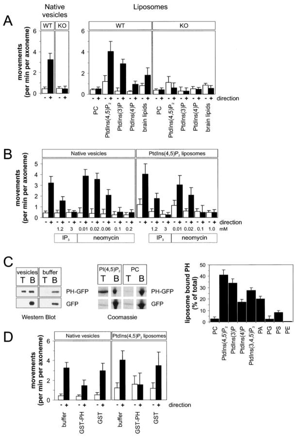
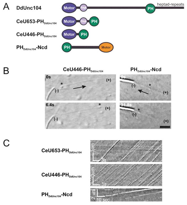
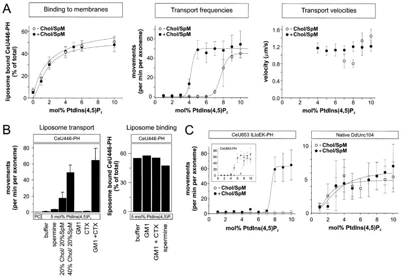
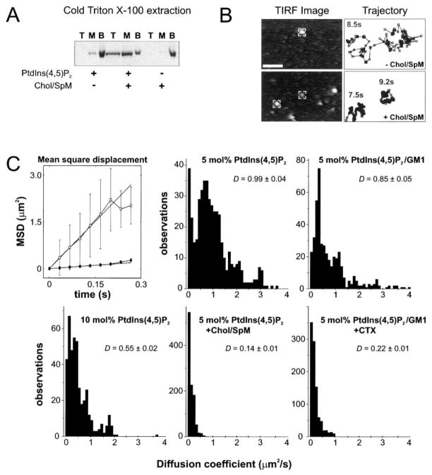
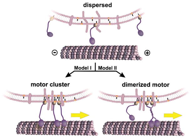
Unc104 (KIF1A) kinesin transports membrane vesicles along microtubules in lower and higher eukaryotes. Using an in vitro motility assay, we show that Unc104 uses a lipid binding pleckstrin homology (PH) domain to dock onto membrane cargo. Through its PH domain, Unc104 can transport phosphatidylinositol(4,5)bisphosphate (PtdIns(4,5)P2)-containing liposomes with similar properties to native vesicles. Interestingly, liposome movement by monomeric Unc104 motors shows a very steep dependence on PtdIns(4,5)P2 concentration (Hill coefficient of approximately 20), even though liposome binding is noncooperative. This switch-like transition for movement can be shifted to lower PtdIns(4,5)P2 concentrations by the addition of cholesterol/sphingomyelin or GM1 ganglioside/cholera toxin, conditions that produce raft-like behavior of Unc104 bound to lipid bilayers. These studies suggest that clustering of Unc104 in PtdIns(4,5)P2-containing rafts provides a trigger for membrane transport.


Figures

Figure 1
Figure 1. The PHDdUnc104 Domain Binds to Membranes and Enables DdUnc104 to Transport Native Vesicles and Artificial Liposomes In Vitro
(A) Motility of liposomes and vesicles in an in vitro assay produced by a DdUnc104-containing motor fraction from wild-type or DdUnc104-knockout (KO) cells. PtdIns(4,5)P2, PtdIns(3)P, and PtdIns(4)P were added at 10 mol% fraction with the remainder being PC. The brain lipids were a mixture of 10% phosphoinositides, 50% phosphatidylserine, and several other brain lipids. The number of membranes moving per min per axoneme were scored. Values show the means and standard deviations (SD) of 3–8 independent experiments. Open bars show minus-end-directed movements; closed bars show plus-end-directed movements. (B) Inhibition of plus-end-directed liposome and vesicle transport (produced by a DdUnc104-containing motor fraction from wild-type cells) by neomycin or soluble inositol(1,4,5)P3 (IP3). Values show the means and SD of four independent experiments. (C) Binding of bacterially-expressed PHDdunc104-GFP, or GFP alone, to native vesicles or liposomes using a membrane floatation assay (Experimental Procedures). After a centrifugation step, membranes were recovered from the top (T) of sucrose step gradient while unbound protein remained at the bottom (B). The left gel (Western blot with an anti-GFP antibody) reveals recovery of PHDdUnc104-GFP in the top fraction only in the presence of vesicles and not in the buffer control. GFP was not recovered in the top fraction in the presence or absence of vesicles. The next image, a Coomassie-stained gel, reveals that PHDdUnc104-GFP binds to 10 mol% PtdIns(4,5)P2 liposomes but not liposomes composed only of PC. Right graph shows quantification of PHDdUnc104-GFP binding to liposomes of various compositions (all at 10 mol% fraction). Values show the mean and SD of three independent experiments. (D) Inhibition of plus-end- but not minus-end-directed transport by addition of bacterially-expressed GST-PHDdunc104 domain (0.24 mg/ml). GST alone added at 0.3 mg/ml had little effect. The in vitro motility assays used a DdUnc104-containing motor fraction from wild-type cells and either 10 mol% PtdIns(4,5)P2 liposomes or native vesicles. Values are the mean and SD of three measurements.
Figure 2
Figure 2. The PHDdUnc104, Joined onto a Kinesin Motor Domain, Elicits Vesicle and Liposome Transport In Vitro
(A) Schematic diagram of native DdUnc104 or minimotor constructs. The PH domain from DdUnc104 was fused C-terminal to the FHA domains (CeU653-PHDdUnc104) or to the motor/neck domain (CeU446-PHDdUnc104) of C. elegans Unc104. For the minus-end-directed, C-terminal motor Ncd, the DdUnc104 PH domain was fused at the N terminus of its elongate coiled-coil domain. See the Experimental Procedure for the precise fusion sites. (B) DIC images show membrane movement (native vesicles indicated by an asterisk) being transported by CeU446-PHDdUnc104 toward the microtubule plus end (+) or by PHDdUnc104-Ncd toward the minus end (−). The axoneme (visible as the higher-contrast elongate object) has longer microtubules growing off of its plus end and shorter microtubules growing off of its minus end (not shown) as a marker of polarity. The scale bar equals 3 μm. (C) Kymographs (see Experimental Procedures) demonstrate the processivity, velocity, and frequency of transport of PtdIns(4,5)P2 liposomes by minimotors. Movement of individual liposome is seen in this plot of the video data as continuous diagonal lines. Note the frequent liposome movement and the steady velocities indicated by parallel straight lines.
Figure 3
Figure 3. Effects of PtdIns(4,5)P2 Concentration and Lipid Clustering Agents on Unc104-Mediated Liposome Transport
(A) The effects of PtdIns(4,5)P2 concentration on motor (CeU446-PHDdUnc104) binding to liposomes (left), frequency of liposome movement (middle), and liposome transport velocity (right). Liposomes were prepared with the indicated concentration of PtdIns(4,5)P2 without (open circle) or with 40 mol% cholesterol/20 mol% sphingomyelin (closed circles), with the remainder being PC. The motor concentration was held fixed at 0.2 μM. Motor binding to liposomes with increasing PtdIns(4,5)P2 concentration showed little or no cooperativity (curves fitted with Hill coefficients of 1.5 ± 0.5 and 0.9 ± 0.7 in the absence and presence of cholesterol/sphingomyelin), while a high degree of cooperativity was observed for liposome transport (curves fitted with Hill coefficients of 20.1 ± 1.0 and 19.8 ± 8.7 in the absence and presence of cholesterol/sphingomyelin). Velocity was scored only when a minimum of ten events could be measured. The mean and SD from four independent experiments are shown. (B) The effects of two lipid clustering agents, cholera toxin (CTX) or spermine, on liposome motility and binding. CTX (2 μg/ml) was used to cluster 10 mol% GM1 incorporated into 5 mol% PtdIns(4,5)P2/85 mol% PC liposomes; 2 mM spermine was added to assays with 5 mol% PtdIns(4,5)P2/95 mol% PC liposomes transported by the minimotor CeU446-PHDdUnc104 (0.2 μM). Chol/SpM liposomes were the same as described in (A). A representative binding experiment of CeU446-PHDdUnc104 to liposomes shows little or no increase in the presence of CTX or spermine. (C) Left: The effect of a destabilized coiled-coil region on liposome transport. CeU653-PHDdUnc104 with the mutations I362E and L365K is indicated, and the behavior of wild-type CeU653-PHDdUnc104 is shown in the insert. Right: Transport of liposomes by a microtubule affinity-purified fraction containing native dimeric DdUnc104 transporting liposomes. The conditions were the same as those described in (A). The means and SD from three independent experiments are shown.
Figure 4
Figure 4. Raft-like Behavior of Unc104 Motor Bound to Liposomes or Supported Lipid Bilayers
(A) 1% Triton X-100 (4°C) extraction of liposomes with bound CeU446-PHDdUnc104 as a biochemical assay of raft formation. The detergent extract was loaded at the bottom of a sucrose gradient, and after centrifugation, top, middle, and bottom (load) fractions were collected and analyzed by SDS-PAGE/Coomassie staining. The minimotor was recovered from detergent-resistant membrane top fraction only when both 5 mol% PtdIns(4,5)P2 and 40 mol% cholesterol/20 mol% sphingomyelin were present in liposomes. (B) Visualization and tracking of single Cy3-labeled CeU446-PHDdUnc104 molecules bound to supported lipid bilayers containing 5 mol% PtdIns(4,5)P2 with or without 40 mol% cholesterol/20 mol% sphingomyelin (remainder PC). The left images show membrane bound motors as visualized by TIRF microscopy. The centroid positions of the “marked” fluorescent molecules were tracked using an automated procedure (Experimental Procedures); the centroid positions measured at 33 ms intervals are shown in the trajectory plots on the right (total time of measurement indicated). These typical plots reveal the restricted motion in the bilayers composed of 5 mol%Pt-dIns(4,5)P2/40 mol% cholesterol/20 mol% sphingomyelin. Very few fluorescent spots were observed on bilayers composed of 100% PC, indicating that binding was specific (not shown). (C) Diffusion coefficients measured on supported membrane bilayers of different lipid coefficients. Examples of mean-square displacement plots calculated from single-molecule trajectories are shown in the absence (open) or presence (closed) of cholesterol/sphingomyelin. Diffusion coefficients (D) were measured as described in the Experimental Procedures; the histograms and the mean ± SD of the mean in μm2/s are shown. The lipid compositions and cholera toxin (CTX) concentration were the same as in Figure 3.
Figure 5
Figure 5. Models for the Regulation of Unc104-Mediated Membrane Transport by PtdIns(4,5)P2 Lipid Clustering
A membrane cargo containing PtdIns(4,5)P2 lipids (orange head groups), transmembrane proteins (light blue), and bound Unc104 motors (dark blue) is shown. In the absence of sufficient PtdIns(4,5)P2 density or clustering, Unc104 motors are dispersed. Since the Unc104 motor is not processive, individual motors transiently interact with the microtubule, but this is insufficient to move the vesicle continuously along the microtubule. PtdIns(4,5)P2 organization into membrane rafts could activate transport by two potential mechanisms. (Model I) Several Unc104 monomeric motors are brought together in a cluster and acting together can maintain attachment to the microtubule and initiate transport. (Model II) Unc104 is a monomer in solution, but the concentration of several motors in a cluster may cause dimerization via predicted coiled-coil regions adjacent to the motor domain. A single, dimerized motor then could move along the microtubule by a hand-over-hand mechanism and processively transport the vesicle.

Similar articles

Cited by

References

    1. Berg JS, Derfler BH, Pennisi CM, Corey DP, Cheney RE. Myosin-X, a novel myosin with pleckstrin homology domains, associates with regions of dynamic actin. J Cell Sci. 2000;113:3439–3451. - PubMed
    1. Bloom GS. The UNC-104/KIF1 family of kinesins. Curr Opin Cell Biol. 2001;13:36–40. - PubMed
    1. Caroni P. Actin cytoskeleton regulation through modulation of PI(4,5)P(2) rafts. EMBO J. 2001;20:4332–4336. - PMC - PubMed
    1. Case RB, Pierce DW, Hom-Booher N, Hart CL, Vale RD. The directional preference of kinesin motors is specified by an element outside of the motor catalytic domain. Cell. 1997;90:959–966. - PubMed
    1. Cohn RC, Poncz L, Waller RL, Dearborn DG. Phosphoinositide content of erythrocyte membranes in cystic fibrosis. J Lab Clin Med. 1988;111:336–340. - PubMed

Publication types

MeSH terms