. 2002 Jun;46(6):1896-905.
doi: 10.1128/AAC.46.6.1896-1905.2002.

Emergence of resistant human immunodeficiency virus type 1 in patients receiving fusion inhibitor (T-20) monotherapy

Affiliations

Emergence of resistant human immunodeficiency virus type 1 in patients receiving fusion inhibitor (T-20) monotherapy

Xiping Wei et al. Antimicrob Agents Chemother. 2002 Jun.

Abstract

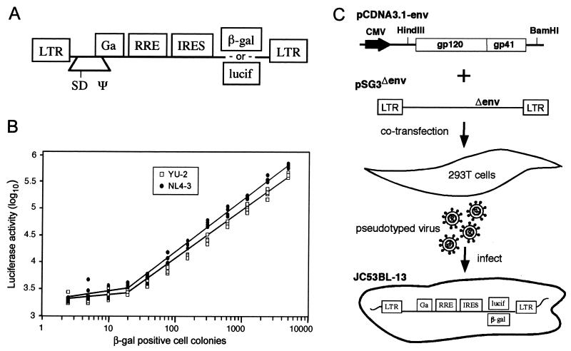
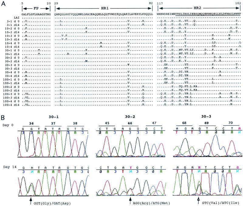
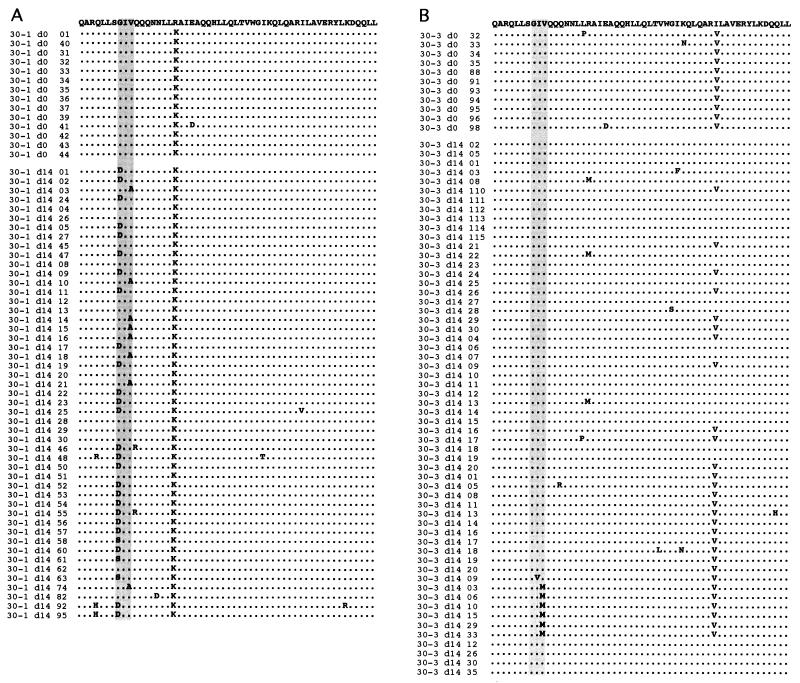
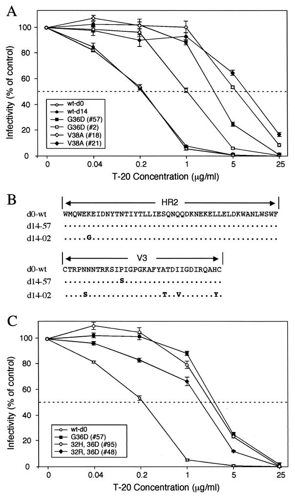
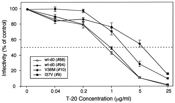
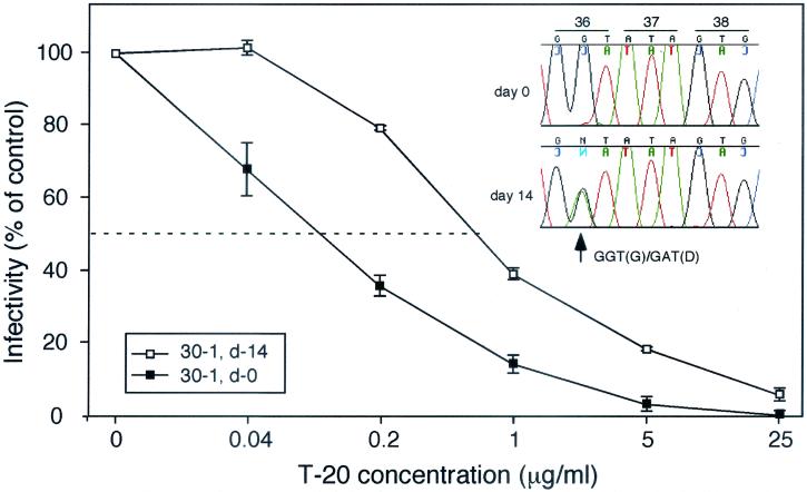
The synthetic peptide T-20 (enfuvirtide) represents the first of a new class of antiretroviral compounds to demonstrate in vivo potency by targeting a step in viral entry. T-20 inhibits a conformational change in the human immunodeficiency virus type 1 (HIV-1) transmembrane glycoprotein (gp41) that is required for fusion between HIV-1 and target cell membranes. The initial phase I clinical trial of T-20 treatment for HIV-infected patients thus provided a unique opportunity to evaluate the emergence of resistant virus in vivo to this novel class of antiretroviral agents. All four patients who received an intermediate dose of T-20 (30 mg twice daily) had an initial decline in plasma viral load over the first 10 days but a rising trend by day 14, suggestive of selection for resistant virus. Plasma virus derived from patients enrolled in all dosage groups of the phase I T-20 trial was analyzed by population sequencing before and after treatment. While no mutations were found within a highly conserved 3-amino-acid sequence (GIV) known to be critical for fusion at baseline, after 14 days of therapy, virus from one patient in the 30-mg dose group (30-1) developed a mutation in this motif, specifically an aspartic acid (D) substitution for glycine (G) at position 36. Multiple env clones were derived from the plasma virus of all four patients in the 30-mg dosage group. Sequence analysis of 49 clones derived from the plasma of patient 30-1 on day 14 revealed that 25 clones contained the G36D mutation, while 8 contained the V38A mutation. Dual mutations involving G36D and other residues within the HR1 domain were also identified. In 5 of the 49 env clones, other mutations involving residues 32 (Q32R or Q32H) and 39 (Q39R) were found in combination with G36D. Cloned env sequences derived from the plasma virus of subject 30-3 also had single mutations in the GIV sequence (V38M and I37V) detectable following therapy with T-20. The plasma virus from subjects 30-2 and 30-4 did not contain changes within the GIV sequence. To analyze the biological resistance properties of these mutations, we developed a novel single-cycle HIV-1 entry assay using JC53BL cells which express beta-galactosidase and luciferase under control of the HIV-1 long terminal repeat. Full-length env clones were derived from the plasma virus of patients 30-1 and 30-3 and used to generate pseudotyped virus stocks. The mean 50% inhibition concentrations (IC(50)s) for mutants G36D and V38A (patient 30-1) were 2.3 microg/ml and 11.2 microg/ml, respectively, statistically significant increases of 9.1- and 45-fold, respectively, compared with those of wild-type Env. The IC(50) for the V38 M mutation (patient 30-3) was 7.6 microg/ml, an 8-fold increase compared with that of the wild type. The I37V mutation resulted in an IC(50) 3.2-fold greater than that of the wild type. Envs with double mutations (Q32R plus G36D and Q32H plus G36D) exhibited a level of resistance similar to that of G36D alone. These findings provide the first evidence for the rapid emergence of clinical resistance to a novel class of HIV-1 entry inhibitors and may be relevant to future treatment strategies involving these agents.


Figures

FIG. 1.
FIG. 1.
HIV-1 entry assay. (A) Illustration of lentivirus vector used to transduce JC53 cells (LTR, long terminal repeat; SD, splice donor; Ψ, RNA packaging signal; RRE, rev-responsive element; IRES, internal ribosomal entry site). Infectious stocks of vector containing either β-Gal or lucif were prepared by transfection of 293T cells as described previously (14) and used to transduce JC53 cells. (B) Analysis of viral infectious units and lucif activity in JC53BL-13 cells. Serial twofold dilutions of the T-tropic NL4-3 and M-tropic YU-2 viruses (1, 11), beginning at 5,120 infectious units, as determined by the β-Gal assay, were used to infect (in quadruplicate) JC53BL-13 cells plated in a 96-well tissue culture plate. Forty-eight hours after infection, the replica wells were lysed, pooled, and analyzed for lucif activity by using an automated Lumistar XL system (BMG Lab Technologies). lucif activity was plotted against the number of infectious units. (C) Phenotypic analysis of the HIV-1 env clones for susceptibility to inhibition by T-20. Full-length env genes were cloned into the pCDNA3.1 eukaryotic expression plasmid. Individual pCDNA3.1-env clones were cotransfected with pSG3Δenv into 293T cells. Pseudotyped progeny virions collected from supernatants of the transfected cells were used to infect JC53BL-13 cells in either the absence or the presence of various concentrations of T-20. Virus infection induces expression of the lucif reporter gene, which was quantified to measure virus entry.
FIG. 2.
FIG. 2.
Population sequence analysis of plasma HIV-1 env. (A) Comparison of the FP, HR1, and HR2 amino acid sequences pre- and posttreatment with T-20. The plasma virus from one subject (3-1) in the 3-mg dose group, two subjects (10-2 and 10-3) in the 10-mg dose group, and four subjects each in the 30- and 100-mg groups were analyzed by population sequencing from before day 0 (d 0) and after 14 days of treatment (d14) with T-20. The sequences were analyzed using Sequencher software and aligned against the LAI consensus sequence. The FP, HR1, and HR2 regions are depicted. The region within HR2 to which T-20 binds is underlined. (B) Quantitative detection of mutant plasma virus by automated population DNA sequencing. DNA sequence chromatograms of specified regions within HR1 are shown for patients 30-1 (residues 36 to 38), 30-2 (residues 45 to 47), and 30-3 (residues 68 to 69). Chromatograms are shown for each subject from before day 0 and after 14 days (Day 14) of T-20 treatment. The sequences shown were obtained from, and therefore are presented as, the minus (noncoding) DNA strand. The minus-strand sequence corresponds to the plus strand (coding), which is indicated on each chromatogram. Codon changes are indicated as plus-strand substitutions.
FIG. 3.
FIG. 3.
Sequence analysis of env clones derived from plasma virus. Prior to (d0) and 14 days after (d14) treatment with T-20, the full-length env gene was amplified from viral RNA by RT-PCR and cloned into pCDNA3.1. The sequences of multiple clones derived from subjects 30-1 (A) and 30-3 (B) were analyzed using Sequencher software and aligned against the LAI consensus sequence. The sequence of the entire HR1 domain is illustrated. The vertical shaded rectangles highlight the GIV motif.
FIG. 4.
FIG. 4.
Sensitivity of GIV mutant Env to inhibition by T-20. One thousand infectious units of HIV-1 pseudotyped with mutant or wt Envs derived from patient 30-1 was used to infect cultures of JC53BL cells containing 0, 0.04, 0.2, 1, 5, and 25 μg of T-20 per ml. Two days later, virus entry was measured by analyzing the indicator cells for lucif activity. Virus infectivity was calculated by dividing the mean lucif activity value at each of the drug concentrations by the mean value of the control (0 mg). Resistance to T-20 is depicted by plotting relative infectivity on the y axis against T-20 concentration on the x axis. (A) HIV-1/SG3Δenv was pseudotyped with the G36D mutant Envs 57 and 2, the V38A mutant Envs 18 and 21, and wt Envs (wt-d0 and wt-d14). (B) Sequence analysis of the HR2 and V3 Env domains for the wt (d0-wt), no. 2 (d14-02), and no. 57 (d14-57) env clones. (C) HIV-1/SG3Δenv stocks were pseudotyped with G36D mutant Env 57, the 32H/36D double mutant Envs (32H, 36D), the 32R/36D double mutant Env (32R, 36D), and the wt Env (wt-d0). The results depicted in panels A and B were highly reproducible and are representative of three independent experiments.
FIG. 5.
FIG. 5.
Sensitivity of GIV mutant Env to inhibition by T-20. HIV-1 pseudotyped with mutant or wt env derived from patient 30-3 was used to infect cultures of JC53BL cells exactly as described for Fig. 4. Pseudotyped HIV-1/SG3Δenv stocks were prepared with the V38M and I37V mutant and wt (wt-d0) Envs. The results are representative of three independent experiments.
FIG. 6.
FIG. 6.
Analysis of T-20 resistance from cultured plasma virus. One thousand infectious units of HIV-1 derived by the coculture of plasma from patient 30-1, prior to (d-0) and after 14 days (d-14) of T-20 treatment, was used to infect JC53BL indicator cells in either the absence or presence of different concentrations (0, 0.04, 0.2, 1, 5, and 25 μg/ml) of T-20. Two days later, the indicator cells were lysed and analyzed for lucif activity. These results are representative of two independent experiments. The DNA sequence chromatogram specific for the GIV sequence is shown (upper right) for the day 14 plasma virus that was isolated after 18 days of cocultivation with normal-donor PBMC.

References

    1. Adachi, A., H. E. Gendelman, S. Koenig, T. Folks, R. Willey, A. Rabson, and M. A. Martin. 1986. Production of acquired immunodeficiency syndrome-associated retrovirus in human and nonhuman cells transfected with an infectious molecular clone. J. Virol. 59:284-291. - PMC - PubMed
    1. Chan, D. C., D. Fass, J. M. Berger, and P. S. Kim. 1997. Core structure of gp41 from the HIV envelope glycoprotein. Cell 89:263-273. - PubMed
    1. Chen, C.-H., T. J. Matthews, C. B. McDanal, D. P. Bolognesi, and M. L. Greenberg. 1995. A molecular clasp in the human immunodeficiency virus (HIV) type 1 TM protein determines the anti-HIV activity of gp41 derivatives: implications for viral fusion. J. Virol. 69:3771-3777. - PMC - PubMed
    1. Condra, J. H., et al. 1995. In vivo emergence of HIV-1 variants resistant to multiple protease inhibitors. Nature 374:569-571. - PubMed
    1. Derdeyn, C. A., J. M. Decker, J. N. Sfakianos, Z. Zhang, W. A. O'Brien, L. Ratner, G. M. Shaw, and E. Hunter. 2001. Sensitivity of human immunodeficiency virus type 1 to fusion inhibitors targeted to the gp41 first heptad repeat involves distinct regions of gp41 and is consistently modulated by gp120 interactions with the coreceptor. J. Virol. 75:8605-8614. - PMC - PubMed

Publication types