. 2002 Aug 9;277(32):28959-71.
doi: 10.1074/jbc.M204525200. Epub 2002 May 17.

Expression cloning and characterization of the C28 acyltransferase of lipid A biosynthesis in Rhizobium leguminosarum

Affiliations

Expression cloning and characterization of the C28 acyltransferase of lipid A biosynthesis in Rhizobium leguminosarum

Shib Sankar Basu et al. J Biol Chem. .

Abstract

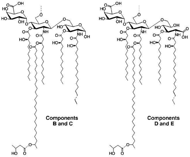
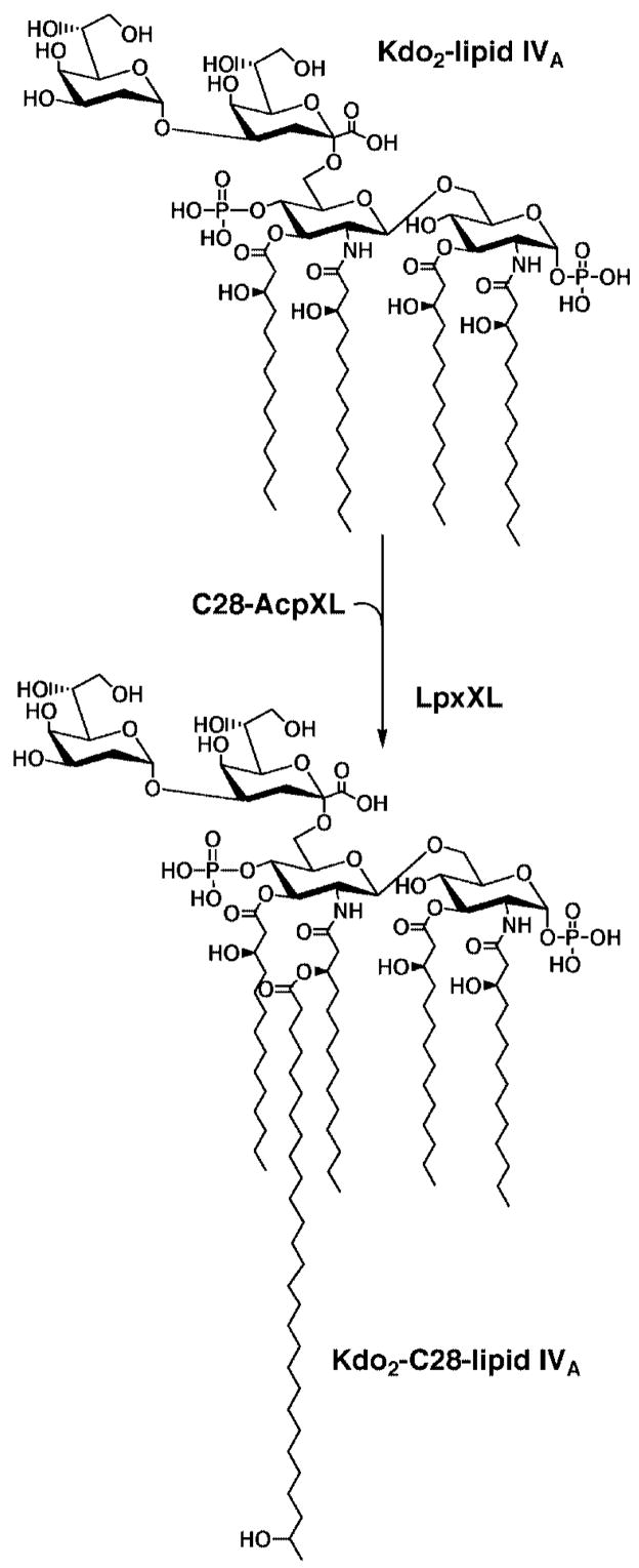
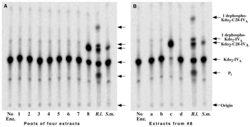
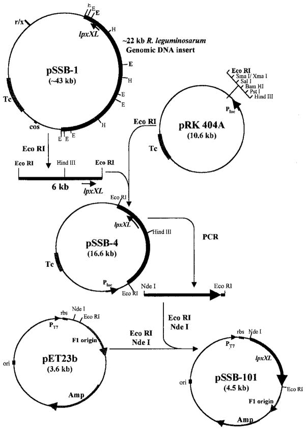
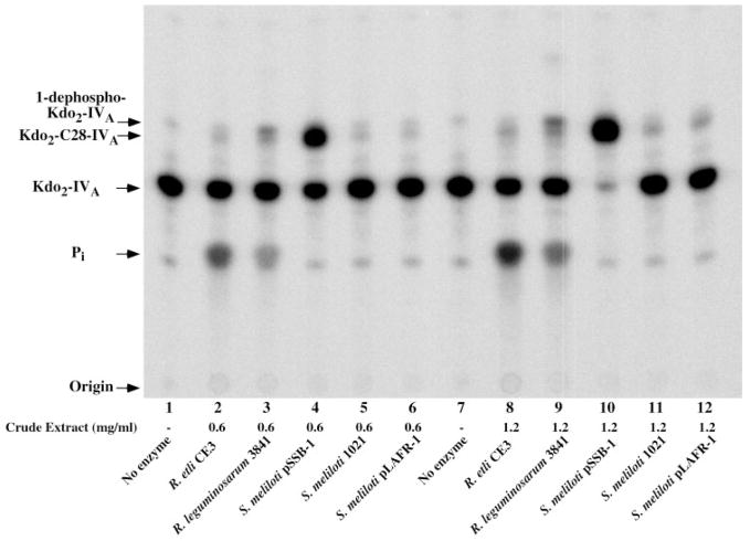
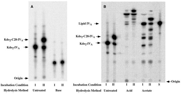
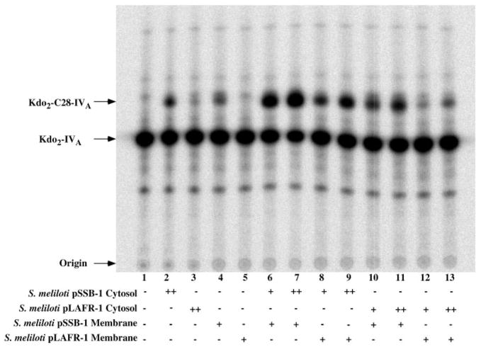
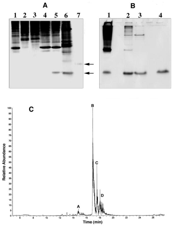
An unusual feature of lipid A from plant endosymbionts of the Rhizobiaceae family is the presence of a 27-hydroxyoctacosanoic acid (C28) moiety. An enzyme that incorporates this acyl chain is present in extracts of Rhizobium leguminosarum, Rhizobium etli, and Sinorhizobium meliloti but not Escherichia coli. The enzyme transfers 27-hydroxyoctacosanate from a specialized acyl carrier protein (AcpXL) to the precursor Kdo2 ((3-deoxy-d-manno-octulosonic acid)2)-lipid IV(A). We now report the identification of five hybrid cosmids that direct the overexpression of this activity by screening approximately 4000 lysates of individual colonies of an R. leguminosarum 3841 genomic DNA library in the host strain S. meliloti 1021. In these heterologous constructs, both the C28 acyltransferase and C28-AcpXL are overproduced. Sequencing of a 9-kb insert from cosmid pSSB-1, which is also present in the other cosmids, shows that acpXL and the lipid A acyltransferase gene (lpxXL) are close to each other but not contiguous. Nine other open reading frames around lpxXL were also sequenced. Four of them encode orthologues of fatty acid and/or polyketide biosynthetic enzymes. AcpXL purified from S. meliloti expressing pSSB-1 is fully acylated, mainly with 27-hydroxyoctacosanoate. Expression of lpxXL in E. coli behind a T7 promoter results in overproduction in vitro of the expected R. leguminosarum acyltransferase, which is C28-AcpXL-dependent and utilizes (3-deoxy-d-manno-octulosonic acid)2-lipid IV(A) as the acceptor. These findings confirm that lpxXL is the structural gene for the C28 acyltransferase. LpxXL is distantly related to the lauroyltransferase (LpxL) of E. coli lipid A biosynthesis, but highly significant LpxXL orthologues are present in Agrobacterium tumefaciens, Brucella melitensis, and all sequenced strains of Rhizobium, consistent with the occurrence of long secondary acyl chains in the lipid A molecules of these organisms.


Figures

Fig. 1
Fig. 1. Structures of the major lipid A molecular species present in R. etli CE3
Components C and E are 3-O-deacylated derivatives of components B and D, respectively (2, 3).
Fig. 2
Fig. 2. Proposed acylation reaction catalyzed by LpxXL
The seven enzymes that make Kdo2-lipid IVA in E. coli are also present in extracts of R. leguminosarum and R. etli (7), and the genes encoding them are found in single copy in the genomes of S. meliloti and M. loti (15, 47). In the LpxXL-catalyzed reaction, acylated AcpXL (but not AcpP) (10) serves as the acyl donor, supplying the 27-hydroxyoctacosanate moiety or the homologous C26 or C30 acyl chains if present. The attachment site for the 27-hydroxyoctacosanate chain generated enzymatically in vitro with Kdo2-lipid IVA as the acceptor has not yet been confirmed, but its attachment is Kdo-dependent (10). However, the proposed location of the 27-hydroxyoctacosanate chain is well established in the lipid A species isolated from cells of R. etli (2, 3).
Fig. 3
Fig. 3. Expression cloning of lpxXL from an R. leguminosarum DNA library in an S. meliloti host
A shows an image of a thin layer plate from the initial screening assay, demonstrating a pool of four lysozyme/EDTA extracts that overexpresses Kdo2-[4′-32P]lipid IVA acylation activity (lane 8). The lane on the far left is the no enzyme control (No Enz.). The lanes designated R.l. are controls in which R. leguminosarum 3841 cell extract prepared by passage through a French pressure cell was used as the enzyme source, and the lanes labeled S.m. are similar S. meliloti 1021 control extracts. B, the four individual cell lysates (labeled a–d) that were used to make pool 8 in A were re-assayed, revealing that only extract c overexpresses the acyltransferase activity. The lane on the left is the no enzyme control (No Enz.). The position of the reaction product generated by the long chain (C28) acyltransferase, which is present at low levels in wild-type R. leguminosarum and S. meliloti extracts (9), is indicated on the right, along with the 1-phosphatase products. The 1-phosphatase is present only in R. leguminosarum extracts (9). The Pi is released by the 4′-phosphatase of R. leguminosarum (9, 11), which likewise is absent in S. meliloti.
Fig. 4
Fig. 4. Construction of plasmids pSSB-4 and pSSB-101 from pSSB-1
The 6-kb EcoRI fragment of pSSB-1 was transferred into the broad range host vector, pRK404A (26), to generate pSSB-4. This construct was first transformed into E. coli HB101, verified by restriction mapping, and then transferred into S. meliloti 1021 by tri-parental mating. Similar procedures were used to construct pSSB-2, pSSB-3, and pSSB-5 (see Fig. 10). After sequencing of pSSB-1, a PCR product containing lpxXL (~960 bp in length) with appropriate restriction was ligated into the expression vector pET23b to generate pSSB-101, which was then transformed into competent cells of E. coli BLR(DE3)/pLysS (Novagen).
Fig. 5
Fig. 5. The pSSB-1-driven overexpression of C28 acyltransferase activity in cell extracts of S. meliloti
Kdo2-[4′-32P]lipid IVA acyltransferase activity in cell extracts of S. meliloti 1021/pSSB-1 was compared with that of wild-type R. leguminosarum, R. etli, S. meliloti 1021, or S. meliloti 1021 harboring the pLAFR-1 cosmid (the vector control). Reactions were carried out in 50 mM Hepes, pH 8.2, 0.2% Triton X-100, and 10μM Kdo2-[4′-32P]lipid IVA, with either 0.6 (lanes 2– 6) or 1.2 mg/ml (lanes 8 –12) of extract protein. After incubation for 10 min at 30 °C, 4-μl portions of each reaction were spotted onto Silica Gel 60 thin layer chromatography plate, which was developed in chloroform/pyridine/88% formic acid/water (30:70:16:10, v/v) and analyzed with a PhosphorImager.
Fig. 6
Fig. 6. Hydrolysis of the LpxXL reaction product with base, acid, or sodium acetate buffer
Incubation I is a control with authentic Kdo2-[4′-32P]lipid IVA. Incubation II shows the behavior of the product generated from Kdo2-[4′-32P]lipid IVA with the S. meliloti 1021/pSSB-1 cell extract. Portions of incubations I and II were hydrolyzed with either base (NaOH), acid (HCl), or pH 4.5 sodium acetate buffer, as described under “Experimental Procedures.” The products of these chemical treatments were analyzed by TLC, followed by PhosphorImager analysis. Panels A and B show results of separate experiments. S is authentic [4′-32P]lipid IVA.
Fig. 7
Fig. 7. Overexpression of both LpxXL and AcpXL in S. meliloti cells harboring pSSB-1
Membrane-free cytosol fractions, prepared from S. meliloti 1021/pSSB-1 or from the vector control S. meliloti 1021/pLAFR-1, were assayed at 0.1 (+) or 0.2 (++) mg/ml protein. Washed membranes were assayed at 0.2 (+) mg/ml protein. The reaction mixtures were incubated at 30 °C for 10 min under standard conditions in the indicated combinations, and 4-μl portions were then spotted onto a silica gel thin layer plate to stop the reactions. Following chromatography, the plate was analyzed with a PhosphorImager.
Fig. 8
Fig. 8. Purification and microcapillary HPLC analysis of acyl-AcpXL from S. meliloti 1021/pSSB-1
A, this gel demonstrates overproduction of acyl-AcpXL in the cytosol of S. meliloti 1021/pSSB-1. Native PAGE was carried out as described under “Experimental Procedures” with 20 μg of cytosolic protein per lane followed by staining with Coomassie Blue; lane 1, S. meliloti 1021; lane 2, R. leguminosarum 3841; lane 3, R. etli CE3; lane 4, S. meliloti 1021; lane 5, S. meliloti 1021/pLAFR-1 (vector control); lane 6, S. meliloti 1021/pSSB-1; lane 7, E. coli AcpP. Lower arrow indicates acyl-AcpXL, and upper arrow E. coli AcpP. B, samples at different stages of the purification were analyzed by SDS-PAGE with 20 μg of protein per lane; lane 1, membrane-free cytosol of S. meliloti 1021/pSSB-1; lane 2, pooled active fractions from DEAE-Sepharose column; lane 3, pooled active fractions from Source-Q15 column; lane 4, pooled active fractions from Superdex-200 column. C, a sample of the purified acyl-AcpXL from S. meliloti 1021/pSSB-1 was fractionated on a microcapillary column packed with Porose 10R2. The column was interfaced with a Finnigan TSQ7000 triple quadrupole mass spectrometer for electrospray ionization mass spectrometry (29). Peaks A–D were analyzed as described in the text.
Fig. 9
Fig. 9. Mass spectrometry of the major acyl-AcpXL component isolated from S. meliloti 1021/pSSB-1
A shows the mass spectrum of the major AcpXL component resolved by microcapillary HPLC (peak B in Fig. 8C). B is the deconvulated data from A, indicating the molecular weights of the two related acyl-AcpXL species present in peak B (Fig. 8C), differing by the presence or absence of the N-terminal methionine residue.
Fig. 10
Fig. 10. Order of genes in the acpXL-lpxXL region of the R. leguminosarum chromosome
Fragments of the ~22-kb DNA insert present in pSSB-1 were subcloned to make pSSB-2, pSSB-3, pSSB-4, and pSSB-5, all of which were sequenced. The dotted lines in the upper part of the figure indicate the ends of the sequenced regions of the insert in pSSB-1, in which there are 11 putative open reading frames that are transcribed in the indicated directions. The lengths of the inserts and the genes are drawn approximately to scale. Similar gene clusters extending from AcpXL to LpxXL are present in the sequenced genomes of A. tumefaciens, S. meliloti, M. loti, and B. melitensis (, –48).
Fig. 11
Fig. 11. Sequence alignments of LpxXL with distantly related late acyltransferases of lipid A biosynthesis
The following abbreviations are used. LpxL_E.coli, E. coli lauroyltransferase involved in lipid A biosynthesis (GenBank accession number AAC74138); Lpx-P_E.coli, E. coli palmitoleoyltransferase involved in lipid A biosynthesis (GenBank accession number AAC75437); LpxM_E.coli, E. coli myristoyltransferase involved in lipid A biosynthesis (GenBank accession number AAC74925); Rickettsia_p, putative lipid A late acyltransferase from R. prowazekii (GenBank accession number CAA15149); LpxXL_R_legu, lipid A acyltransferase A described here; Chlamydia_tr, putative lipid A acyltransferase from Chlamydia trachomatis (Gen-Bank accession number AAC67600). Conserved amino acids are colored. The identical residues noted in yellow may form part of the active site. The C-terminal 122 residue extension of the C. trachomatis protein is not shown.
Fig. 12
Fig. 12. Expression and assay of LpxXL in E. coli membranes
A, SDS-PAGE of membrane proteins, prepared from isopropyl-1-thio-β-D-galactopyranoside-induced E. coli cells harboring either pSSB-101 or the vector control pET23b, was carried out with 20-μg samples of membrane protein per lane. These were run beside molecular weight standards, as indicated. B, the assays of recombinant LpxXL activity in E. coli membranes were performed in the presence or absence of 0.05 mg/ml purified AcpXL from S. meliloti 1021/pSSB-1, as indicated. Washed membranes from the pET23 vector control or the lpxXL expressing strain E. coli/pSSB-101 were added at 10 or 20 μg/ml. The reaction mixtures were incubated for 5 min at 30 °C, as described under “Experimental Procedures.”

References

    1. Raetz CRH, Whitfield C. Annu Rev Biochem. 2002;71:635–700. - PMC - PubMed
    1. Que NLS, Lin S, Cotter RJ, Raetz CRH. J Biol Chem. 2000;275:28006–28016. - PMC - PubMed
    1. Que NLS, Ribeiro AA, Raetz CRH. J Biol Chem. 2000;275:28017–28027. - PMC - PubMed
    1. Bhat UR, Forsberg LS, Carlson RW. J Biol Chem. 1994;269:14402–14410. - PubMed
    1. Bhat UR, Mayer H, Yokota A, Hollingsworth RI, Carlson R. J Bacteriol. 1991;173:2155–2159. - PMC - PubMed

Publication types

MeSH terms

Associated data

LinkOut - more resources