Comparative Study
. 2002 May 15;22(10):4080-94.
doi: 10.1523/JNEUROSCI.22-10-04080.2002.

Age and sex differences in mitotic activity within the zebra finch telencephalon

Affiliations
Comparative Study

Age and sex differences in mitotic activity within the zebra finch telencephalon

Valerie DeWulf et al. J Neurosci. .

Abstract

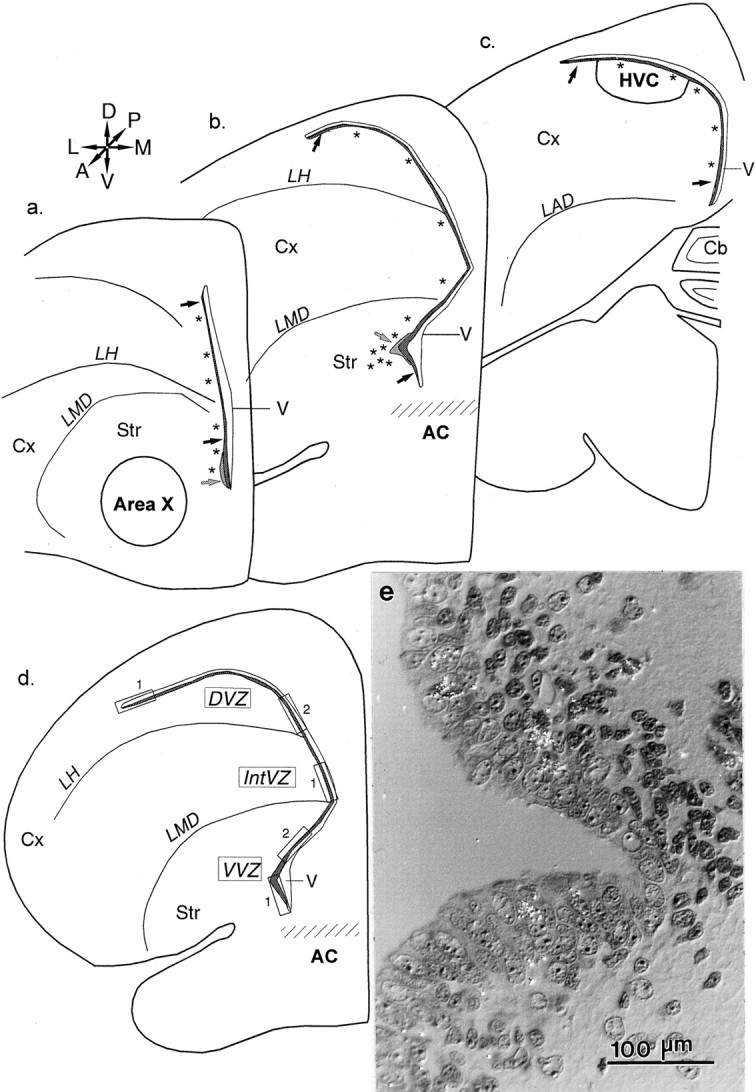
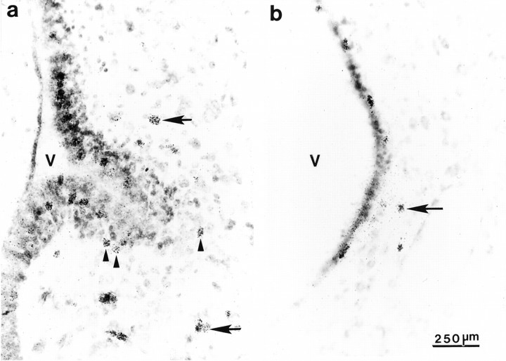
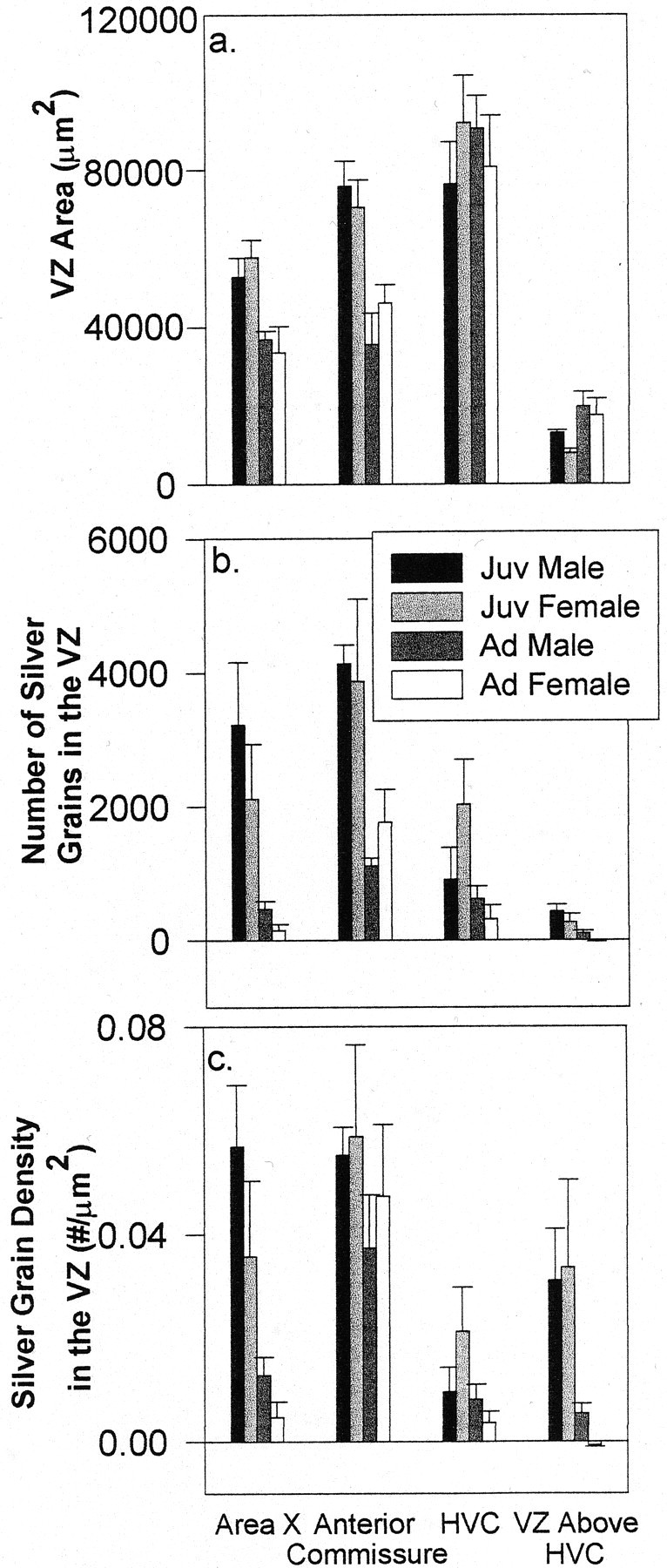
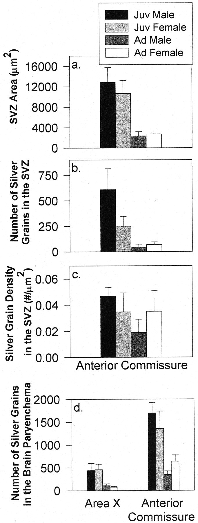
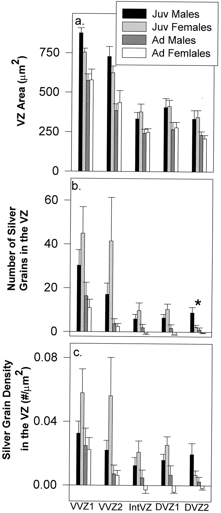
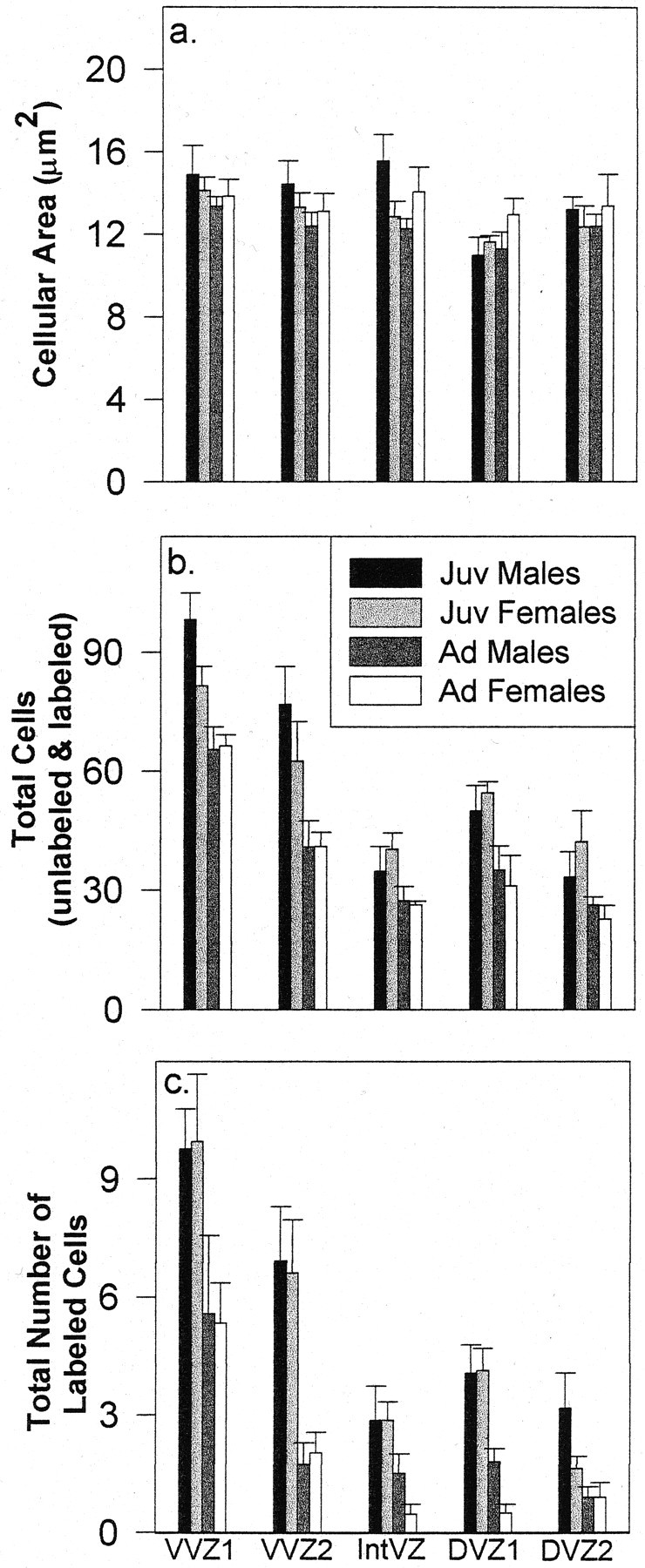
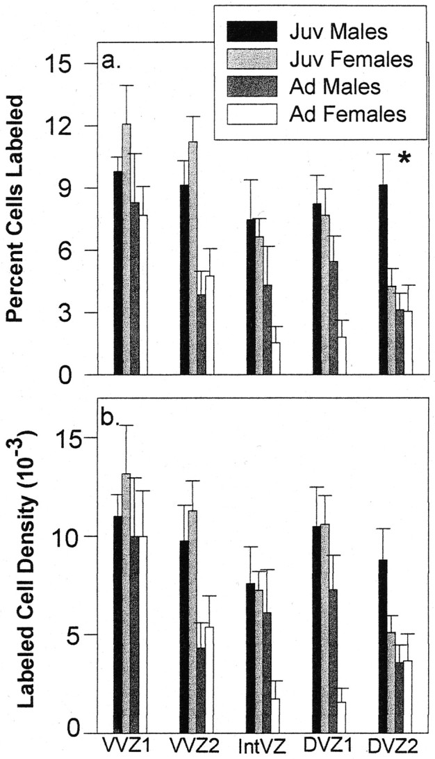
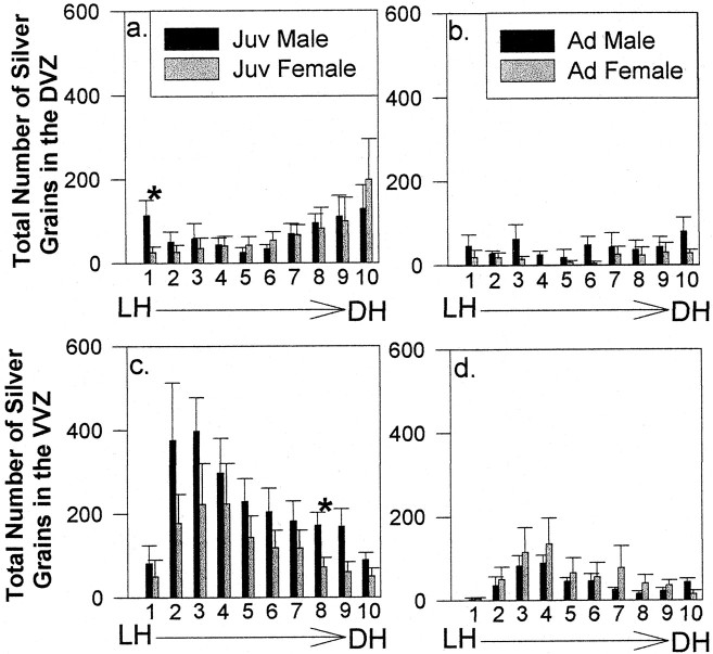
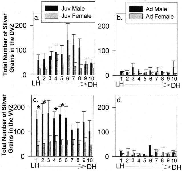
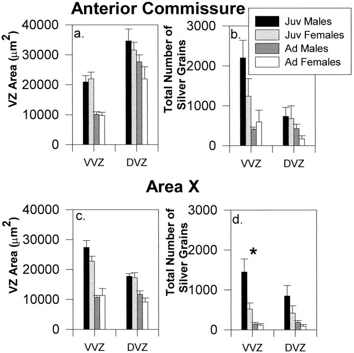
Brain regions associated with song learning in zebra finches are larger and contain more neurons in males than females. Differences in cell proliferation, migration, survival, and specification may all contribute to the divergent development of the song-control system in developing birds. This study quantified levels of cell proliferation within the telencephalic ventricular zone (VZ) of juvenile and adult birds to look for both age and sex differences in mitotic activity that might contribute to the construction of song-control circuits. A single pulse of [(3)H]thymidine was administered to juveniles and adults of both sexes, and animals were killed 2 hr later. Analysis of thymidine labeling within the telencephalic VZ at the levels of area X, the anterior commissure, and high vocal center (HVC) revealed two major findings: (1) levels of mitotic activity decreased as a function of age in both males and females because of a reduction in the number of dividing cells within the VZ, and (2) sex differences in thymidine labeling occurred in restricted, localized segments of the VZ at the levels of area X and the anterior commissure in juveniles but not adults. Thus, overall proliferative activity decreases as birds mature, and the incidence of cell division in all regions of the VZ becomes equivalent in both sexes, such that no regions of sexually dimorphic proliferation are evident by adulthood. These data suggest that regions of sexually dimorphic proliferation within the VZ may contain precursor cells that give rise to song-control neurons, such that higher rates of mitotic activity in juvenile males could contribute to the growth of song-control nuclei such as HVC and area X.


Figures

Fig. 1.
Fig. 1.
Schematic coronal sections through the zebra finch brain at the levels of area X (a), the anterior commissure (b), and HVC (c). A, Anterior;D, dorsal; L, lateral; M, medial; P, posterior, V, ventral. Proliferation in the telencephalon occurs in the epithelium adjacent to the lateral ventricle, the VZ (black arrows), and the SVZ (gray arrows). Asterisksindicate the locations of thymidine-labeled cell clusters in the brain parenchyma. d, A separate cross section of the telencephalon indicating the boxed regions measured in the 1 μm plastic tissue analysis. e, Photomicrograph of the VZ in a 1-μm-thick section demonstrating the out-pocketing of the VZ in juvenile birds. Cx, Cortex;Str, striatum; Cb, cerebellum;V, lateral ventricle; LAD, lamina archistriatalis dorsalis; AC, anterior commissure;LH, lamina hyperstriatica; LMD, lamina medullaris dorsalis.
Fig. 2.
Fig. 2.
Autoradiograms showing thymidine labeling within the ventral portion of the VZ at the level of the anterior commissure in a juvenile male (a) and an adult female (b). Thymidine labeling in the SVZ (arrowheads) and clusters of thymidine-labeled cells (arrows) adjacent to the VZ are apparent. A large out-pocketing in the VZ is apparent in juveniles, whereas the out-pocketing is much reduced in adults. V, Lateral ventricle.
Fig. 3.
Fig. 3.
A 10 μm tissue analysis demonstrating the area of the VZ (a), the total number of silver grains (b), and silver-grain density within the VZ (c) at the levels of area X, the anterior commissure, HVC, and the VZ immediately above HVC in juveniles (Juv) and adults (Ad) of both sexes (mean ± SEM).
Fig. 4.
Fig. 4.
A 10 μm tissue analysis demonstrating the area of the SVZ (a), the total number of silver grains (b), and silver-grain density within the SVZ (c) at the level of the anterior commissure in juveniles (Juv) and adults (Ad) (mean ± SEM). d, The total number of silver grains overlying labeled cell clusters adjacent to the VZ within the brain parenchyma in 10-μm-thick tissue at the levels of area X and the anterior commissure in juveniles and adults of both sexes (mean ± SEM).
Fig. 5.
Fig. 5.
A 1 μm tissue analysis demonstrating VZ area (a), the total number of silver grains (b), and silver-grain density (c) (segments shown in Fig. 1d) at the level of the anterior commissure in juveniles (Juv) and adults (Ad) of both sexes (mean ± SEM). Theasterisk indicates a main effect of sex in the total number of silver grains in DVZ2.
Fig. 6.
Fig. 6.
A 1 μm tissue analysis demonstrating cellular area (a), the total number of cells (labeled and unlabeled) (b), and the total number of labeled cells (c) within each VZ segment measured at the level of the anterior commissure in juveniles (Juv) and adults (Ad) of both sexes (mean ± SEM).
Fig. 7.
Fig. 7.
A 1 μm tissue analysis demonstrating the percentage of cells labeled (a) and labeled cell density (b) within each VZ segment measured at the level of the anterior commissure in juveniles (Juv) and adults (Ad) of both sexes (mean ± SEM). Theasterisk indicates a significant difference in the proportion of labeled cells between juvenile males and females in DVZ2.
Fig. 8.
Fig. 8.
Histograms of the total number of silver grains within each bin of the DVZ of juveniles (Juv) (a) and adults (Ad) (b) and in the VVZ of juveniles (c) and adults (d) at the level of the anterior commissure (mean ± SEM). Asterisks indicate significant differences in thymidine labeling between juvenile males and females in DVZ bin 1 (a) and VVZ bin 8 (c). DH, Dorsal horn.
Fig. 9.
Fig. 9.
Histograms of the total number of silver grains within each bin of the DVZ of juveniles (Juv) (a) and adults (Ad) (b) and in the VVZ of juveniles (c) and adults (d) at rostral levels of area X (mean ± SEM). Asterisksin c indicate significant differences in thymidine labeling between juvenile males and females in VVZ bins 1, 2, 4, and 5.DH, Dorsal horn.
Fig. 10.
Fig. 10.
The area and total number of silver grains within the DVZ and VVZ at the levels of the anterior commissure (a, b) and area X (c, d) (mean ± SEM). The asterisk in d indicates a significant difference between juvenile (Juv) males and females within the VVZ at area X. Ad, Adult.

References

    1. Alvarez-Buylla A, Kirn JR. Birth, migration, incorporation, and death of vocal control neurons in adult songbirds. J Neurobiol. 1997;33:585–601. - PubMed
    1. Alvarez-Buylla A, Nottebohm F. Migration of young neurons in adult avian brain. Nature. 1988;335:353–354. - PubMed
    1. Alvarez-Buylla A, Theelen M, Nottebohm F. Birth of projection neurons in the higher vocal center of the canary forebrain before, during, and after song learning. Proc Natl Acad Sci USA. 1988;85:8722–8726. - PMC - PubMed
    1. Alvarez-Buylla A, Theelen M, Nottebohm F. Proliferation “hot spots” in adult avian ventricular zone reveal radial cell division. Neuron. 1990;5:101–109. - PubMed
    1. Alvarez-Buylla A, Ling CY, Nottebohm F. High vocal center growth and its relation to neurogenesis, neuronal replacement and song acquisition in juvenile canaries. J Neurobiol. 1992;23:396–406. - PubMed

Publication types

MeSH terms

LinkOut - more resources