. 2002 May;109(10):1303-10.
doi: 10.1172/JCI14506.

Paracrine regulation of fat cell formation in bone marrow cultures via adiponectin and prostaglandins

Affiliations

Paracrine regulation of fat cell formation in bone marrow cultures via adiponectin and prostaglandins

Takafumi Yokota et al. J Clin Invest. 2002 May.

Abstract

Adiponectin, an adipocyte-derived hormone, was recently shown to have potential therapeutic applications in diabetes and obesity because of its influence on glucose and lipid metabolism. We found that brown fat in normal human bone marrow contains this protein and used marrow-derived preadipocyte lines and long-term cultures to explore potential roles in hematopoiesis. Recombinant adiponectin blocked fat cell formation in long-term bone marrow cultures and inhibited the differentiation of cloned stromal preadipocytes. Adiponectin also caused elevated expression of cyclooxygenase-2 (COX-2) by these stromal cells and induced release of prostaglandin E(2) (PGE(2)). The COX-2 inhibitor Dup-697 prevented the inhibitory action of adiponectin on preadipocyte differentiation, suggesting involvement of stromal cell-derived prostanoids. Furthermore, adiponectin failed to block fat cell generation when bone marrow cells were derived from B6,129S(Ptgs2tm1Jed) (COX-2(+/-)) mice. These observations show that preadipocytes represent direct targets for adiponectin action, establishing a paracrine negative feedback loop for fat regulation. They also link adiponectin to the COX-2-dependent PGs that are critical in this process.


Figures

Figure 1
Figure 1
Adiponectin is present in normal human bone marrow. (a) Total RNA derived from normal human bone marrow was analyzed by RT-PCR. Samples containing all reagents except human bone marrow cDNA were used as negative controls. (b) Normal human bone marrow was processed and stained with a monoclonal antibody to adiponectin or an isotype-matched irrelevant control antibody.
Figure 2
Figure 2
Recombinant adiponectin inhibits adipogenesis in culture. (a) Recombinant adiponectin (right lanes) was subjected to SDS-PAGE under either nonreducing or reducing conditions and stained with Coomassie brilliant blue. Protein size markers are shown for comparison (left lanes). (b) Analytical gel filtration chromatography was performed with recombinant adiponectin. Arrows indicate the apparent molecular weight of each peak. (c) Fat cell formation in adherent layers of Dexter cultures (top and middle panels, at 6 weeks; bottom panel, at 12 weeks from initiation of culture) is shown in these phase-contrast micrographs. Adiponectin was withdrawn after 6 weeks of culture (bottom panel). Arrows in each picture indicate adipocytes. The data is representative of that obtained in three similar experiments.
Figure 3
Figure 3
Adiponectin induces the COX-2–prostanoid pathway in preadipocytes. (a) Northern blot analysis of COX-2 and COX-1 expression in BMS2 cells treated with adiponectin. (b) Adiponectin upregulates PGE2 secretion by BMS2 cells (open circles, BSA; closed circles, adiponectin). The data are presented as the mean of duplicate cultures. Similar results were obtained in four independent experiments.
Figure 4
Figure 4
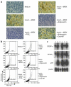
Adiponectin inhibits the differentiation of preadipocytes to adipocytes. Fat cell differentiation from BMS2 cells cultured with the indicated substances for 10 days in 24-well plates is illustrated in phase-contrast photomicrographs (a) and by flow cytometry with Nile red staining (b). The numbers in the boxes indicate the mean ± SD percentages of adipocytes in triplicate cultures. The data are representative of that obtained in three similar experiments. (c) Expression of adipocyte-specific genes (C/EBP-α, PPAR-γ, and aP2) and β-actin in BMS2. Poly(A)+ mRNA was isolated from BMS2 cells cultured under the indicated conditions and subjected to Northern blot analysis. Lane 1, medium alone; lane 2, insulin + MIBX; lane 3, insulin + MIBX + PGE2; lane 4, insulin + MIBX + GST; lane 5, insulin + MIBX + adiponectin; lane 6, insulin + MIBX + adiponectin + Dup-697.
Figure 5
Figure 5
Preadipocytes from COX-2+/– mice are resistant to adiponectin. Fat cells were conspicuous in adherent layers of bone marrow cultures established from heterozygous COX-2+/– mice, as illustrated with these phase-contrast photomicrographs. Similar results were obtained in two independent experiments.

References

    1. Pittenger MF, et al. Multilineage potential of adult human mesenchymal stem cells. Science. 1999;284:143–147. - PubMed
    1. Gimble JM, et al. Adipogenesis in a murine bone marrow stromal cell line capable of supporting B lineage lymphocyte growth and proliferation: biochemical and molecular characterization. Eur J Immunol. 1990;20:379–387. - PubMed
    1. Russell TR, Ho R. Conversion of 3T3 fibroblasts into adipose cells: triggering of differentiation by prostaglandin F2alpha and 1-methyl-3-isobutyl xanthine. Proc Natl Acad Sci USA. 1976;73:4516–4520. - PMC - PubMed
    1. Forman BM, et al. 15-deoxy-delta 12,14-prostaglandin J2 is a ligand for the adipocyte determination factor PPAR gamma. Cell. 1995;83:803–812. - PubMed
    1. Kliewer SA, et al. A prostaglandin J2 metabolite binds peroxisome proliferator-activated receptor gamma and promotes adipocyte differentiation. Cell. 1995;83:813–819. - PubMed

Publication types

MeSH terms