. 2002 May;109(10):1351-9.
doi: 10.1172/JCI14704.

Differential responses of mast cell Toll-like receptors 2 and 4 in allergy and innate immunity

Affiliations

Differential responses of mast cell Toll-like receptors 2 and 4 in allergy and innate immunity

Volaluck Supajatura et al. J Clin Invest. 2002 May.

Abstract

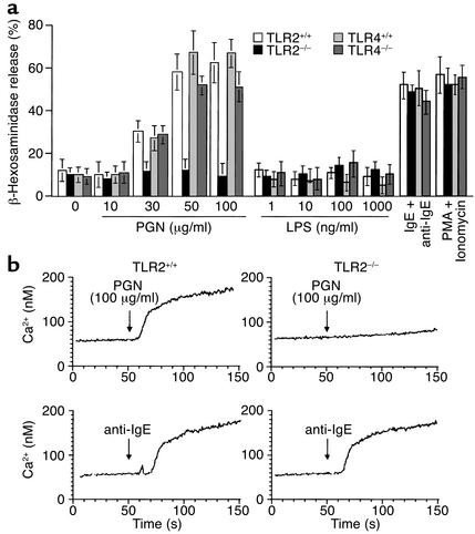
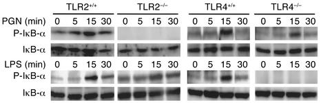
Toll-like receptor 2 (TLR2) and TLR4 play important roles in the early innate immune response to microbial challenge. To clarify the functional roles of TLRs 2 and 4 in mast cells, we examined bone marrow-derived mast cells (BMMCs) from TLR2 or TLR4 gene-targeted mice. Peptidoglycan (PGN) from Staphylococcus aureus stimulated mast cells in a TLR2-dependent manner to produce TNF-alpha, IL-4, IL-5, IL-6, and IL-13, but not IL-1beta. In contrast, LPS from Escherichia coli stimulated mast cells in a TLR4-dependent manner to produce TNF-alpha, IL-1beta, IL-6, and IL-13, but not IL-4 nor IL-5. Furthermore, TLR2- but not TLR4-dependent mast cell stimulation resulted in mast cell degranulation and Ca2+ mobilization. In a mast cell-dependent model of acute sepsis, TLR4 deficiency of BMMCs in mice resulted in significantly higher mortality because of defective neutrophil recruitment and production of proinflammatory cytokines in the peritoneal cavity. Intradermal injection of PGN led to increased vasodilatation and inflammation through TLR2-dependent activation of mast cells in the skin. Taken together, these results suggest that direct activation of mast cells via TLR2 or TLR4 by respective microligands contributes to innate and allergic immune responses.


Figures

Figure 1
Figure 1
PGN but not LPS induces mast cell degranulation and Ca2+ mobilization through TLR2. (a) BMMCs from TLR2+/+ (white bars), TLR2–/– (black bars), TLR4+/+ (light gray bars), and TLR4–/– (dark gray bars) were incubated with indicated concentrations of PGN or LPS. β-Hexosaminidase in the supernatant was measured as described in Methods. Data shown are mean ± SD of three experiments conducted with different BMMC preparations. BMMCs stimulated with IgE and anti-IgE or PMA and ionomycin were used as positive control. (b) TLR2+/+ and TLR2–/– BMMCs were stimulated with 100 μg/ml of PGN; then Ca2+ mobilization was evaluated as described in Methods. BMMCs stimulated with IgE and anti-IgE were used as positive control. Data shown is one representative result from several experiments that had similar results.
Figure 2
Figure 2
PGN-TLR2 stimulation and LPS-TLR4 stimulation lead to different cytokine production by BMMCs. BMMCs from TLR2+/+ (white bars), TLR2–/– (black bars), TLR4+/+ (light gray bars), and TLR4–/– (dark gray bars) were stimulated with indicated concentrations of PGN (a) or LPS (b), 3 hours for TNF-α and 6 hours for other cytokines. The concentrations of cytokine in the supernatant were determined as described in Methods. Data shown are mean ± SD of three experiments conducted with different BMMC preparations.
Figure 3
Figure 3
Stimulation of mast cells via both TLR2 and TLR4 leads to activation of NF-κB signaling. BMMCs were stimulated with 100 μg/ml of PGN or 50 ng/ml of LPS for 5, 15, and 30 minutes. Then lysates were subjected to SDS-PAGE and immunoblotted with antibody to phosphorylated IκB-α (P-IκB-α). The same blots were stripped and reblotted with antibody to IκB-α. The result shown is a representative of three experiments conducted with different BMMC preparations that had similar results.
Figure 4
Figure 4
Mast cell TLR4 but not mast cell TLR2 is required for the full expression of innate immunity in a mast cell–dependent sepsis model. (a) W/Wv mice without reconstitution or 5 weeks after reconstitution with BMMCs from TLR4+/+, TLR4–/–, TLR2+/+, or TLR2–/– mice (6–16 mice per group) underwent CLP. The mortality was observed and expressed as percent survival. W/Wv mice without BMMC reconstitution (injected with saline) were the control group. (b) Six hours after CLP, the mice were killed and peritoneal fluid was investigated. The total numbers of leukocytes and neutrophils were estimated by differential count and expressed as total cells per mouse. (c) The levels of indicated cytokines were measured by ELISA kit. Data shown are mean ± SD of three mice.
Figure 5
Figure 5
TLR2 on mast cell–dependent skin inflammation upon application of PGN. Reconstitution of skin mast cells of W/Wv mice by TLR2–/–, TLR4–/–, or wild-type BMMCs was done as described in Methods. Wild-type mice (a) or indicated mice (b) were intravenously injected with 0.5% Evans blue 5 minutes before intradermal application and 20 μl of PGN (100 μg/ml) or LPS (100 ng/ml) into the left ear or saline into the right ear. The mice were sacrificed 15 minutes after PGN or LPS injection, and dye contents in the tissues were measured as described in Methods. Data shown are mean ± SD of three mice. (c) Reconstitution of skin mast cells of W/Wv mice by TLR2–/–, TLR4–/–, or wild-type BMMCs was histologically confirmed by Alcian blue and Safranin staining (AS; without any treatment; upper panels) and Toluidine blue staining (TB; saline-injected; lower panels). (d) Mice were intradermally injected with PGN (upper four rows) or LPS (bottom row) as described above. Mice were sacrificed 15 minutes (left and middle columns) or 4 hours later (right column), and ear sections from W/Wv (first row), W/Wv/wild-type (W/Wv/WT; second row), W/Wv/TLR4–/– (third row), or W/Wv/TLR2–/– mice (fourth row) were stained with H&E (middle and right columns) or Toluidine blue (TB; left column) for histological evaluations. All magnification is ×600, except the figures at 4 hours after PGN application from W/Wv/WT and W/Wv/TLR4–/– and at 4 hours after LPS application (all ×200).

References

    1. Metcalfe DD, Baram D, Mekori YA. Mast cells. Physiol Rev. 1997;77:1033–1079. - PubMed
    1. Weber S, et al. Mast cells. Int J Dermatol. 1995;34:1–10. - PubMed
    1. Bradding P, Holgate ST. Immunopathology and human mast cell cytokines. Crit Rev Oncol Hematol. 1999;31:119–133. - PubMed
    1. Galli SJ, Wershil BK. The two faces of the mast cells. Nature. 1996;381:21–22. - PubMed
    1. Malaviya R, Ikeda T, Ross E, Abraham SN. Mast cell modulation of neutrophil influx and bacterial clearance at sites of infection through TNF-alpha. Nature. 1996;381:77–80. - PubMed

Publication types

MeSH terms