Clinical Trial
. 2002 May 20;195(10):1279-88.
doi: 10.1084/jem.20012100.

Rapid induction of tumor-specific type 1 T helper cells in metastatic melanoma patients by vaccination with mature, cryopreserved, peptide-loaded monocyte-derived dendritic cells

Affiliations
Clinical Trial

Rapid induction of tumor-specific type 1 T helper cells in metastatic melanoma patients by vaccination with mature, cryopreserved, peptide-loaded monocyte-derived dendritic cells

Beatrice Schuler-Thurner et al. J Exp Med. .

Erratum in

  • J Exp Med. 2003 Feb 3;197(3):395.

Abstract

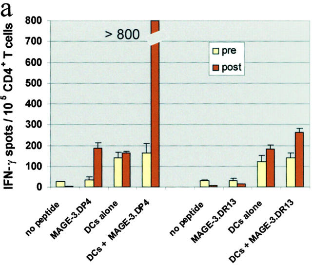
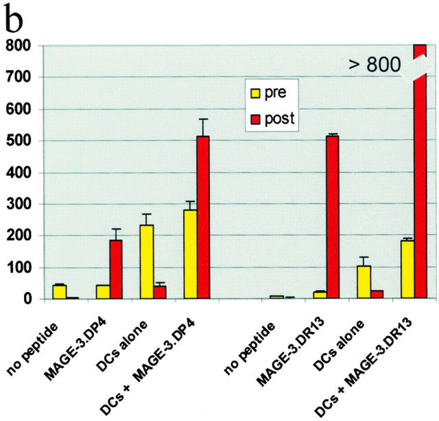
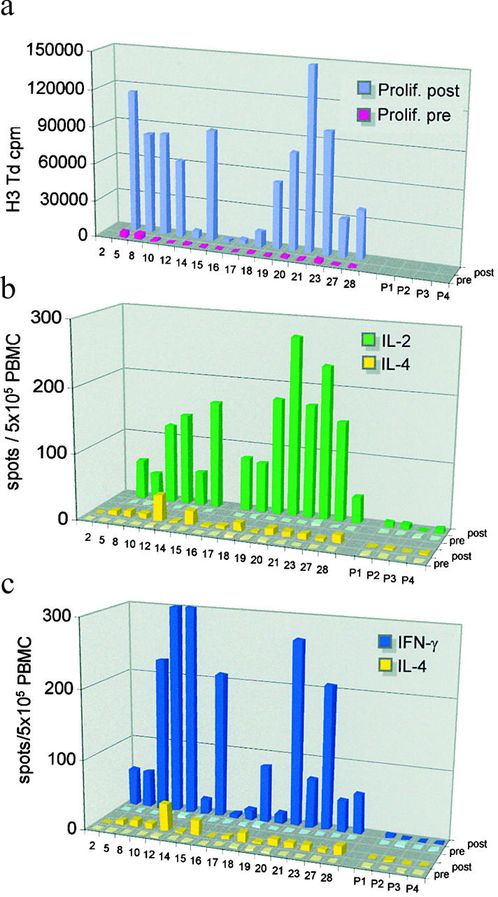
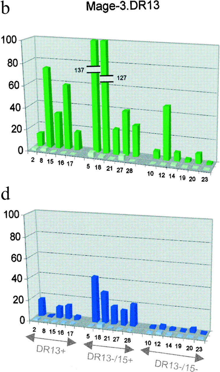
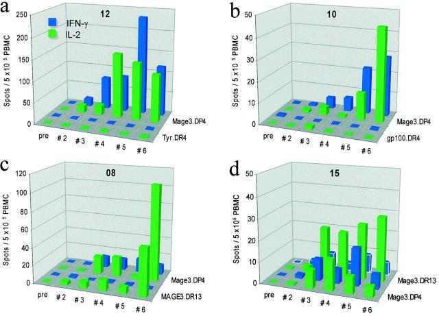
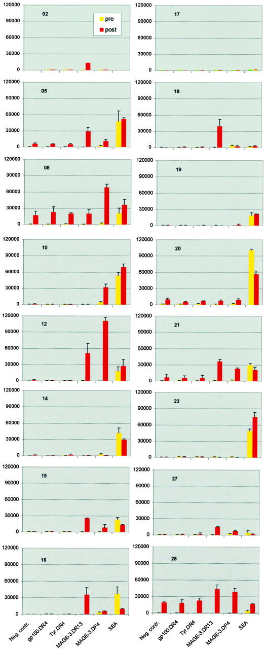
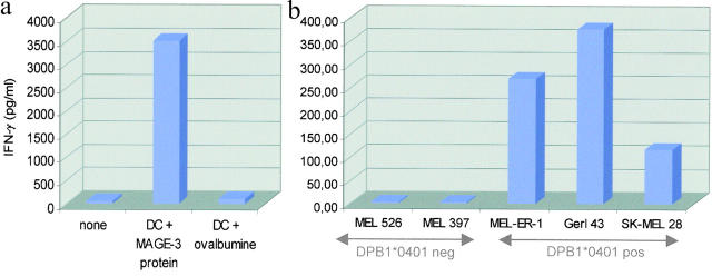
There is consensus that an optimized cancer vaccine will have to induce not only CD8+ cytotoxic but also CD4+ T helper (Th) cells, particularly interferon (IFN)-gamma-producing, type 1 Th cells. The induction of strong, ex vivo detectable type 1 Th cell responses has not been reported to date. We demonstrate now that the subcutaneous injection of cryopreserved, mature, antigen-loaded, monocyte-derived dendritic cells (DCs) rapidly induces unequivocal Th1 responses (ex vivo detectable IFN-gamma-producing effectors as well as proliferating precursors) both to the control antigen KLH and to major histocompatibility complex (MHC) class II-restricted tumor peptides (melanoma-antigen [Mage]-3.DP4 and Mage-3.DR13) in the majority of 16 evaluable patients with metastatic melanoma. These Th1 cells recognized not only peptides, but also DCs loaded with Mage-3 protein, and in case of Mage-3DP4-specific Th1 cells IFN-gamma was released even after direct recognition of viable, Mage-3-expressing HLA-DP4+ melanoma cells. The capacity of DCs to rapidly induce Th1 cells should be valuable to evaluate whether Th1 cells are instrumental in targeting human cancer and chronic infections.


Figures

Figure 6.
Figure 6.
Induction of tumor-specific recall Th1 responses by DC immunization Pre and posttherapy (i.e., 4 wk after vaccination no. 5) CD4+ T cells from patients 12 (a) and 15 (b) were stimulated once in vitro with autologous, peptide-loaded mature DCs (10 μg/ml). On day 8 T cells were stimulated either with peptide alone (Mage-3.DP-4 or Mage-3.DR13 peptide) or with peptide-loaded mature DCs and IFN-γ spot-forming cells were measured by the Elispot technique. The results shown represent the average and SD of triplicate cocultures. Note that Mage-3.DP4-specific and Mage-3.DR13-specific IFN-γ spot-producing CD4+ Th1 cells are clearly expanded after DC vaccination in patients 12 and 15, respectively which fits to the results of the respective proliferative assays (Fig. 5). Note also that (auto-) reactivity to peptide-unloaded DCs (generated in patient 12 from post, and in patient 15 from prevaccination PBMCs) is not increased, but rather decreased upon DC vaccination and induction of specific immunity.
Figure 6.
Figure 6.
Induction of tumor-specific recall Th1 responses by DC immunization Pre and posttherapy (i.e., 4 wk after vaccination no. 5) CD4+ T cells from patients 12 (a) and 15 (b) were stimulated once in vitro with autologous, peptide-loaded mature DCs (10 μg/ml). On day 8 T cells were stimulated either with peptide alone (Mage-3.DP-4 or Mage-3.DR13 peptide) or with peptide-loaded mature DCs and IFN-γ spot-forming cells were measured by the Elispot technique. The results shown represent the average and SD of triplicate cocultures. Note that Mage-3.DP4-specific and Mage-3.DR13-specific IFN-γ spot-producing CD4+ Th1 cells are clearly expanded after DC vaccination in patients 12 and 15, respectively which fits to the results of the respective proliferative assays (Fig. 5). Note also that (auto-) reactivity to peptide-unloaded DCs (generated in patient 12 from post, and in patient 15 from prevaccination PBMCs) is not increased, but rather decreased upon DC vaccination and induction of specific immunity.
Figure 1.
Figure 1.
DTH reactions to peptide-loaded DCs and to Mage-3DP4 peptide alone. (a) Massive local reaction at the vaccination site to peptide-loaded mature DCs in patient 12 (>10 cm induration at 48 h), (b) DTH reaction (arrows) to intradermally administered MAGE-3.DP4 peptide. No DTH after injection of vehicle, MelanA.A2, or Mage-3.DR13 peptides.
Figure 2.
Figure 2.
Priming to KLH after a single injection of KLH-pulsed DCs. (a) Proliferation assay. Pre and postvaccination (i.e., before and 12 wk after the single injection of 4 million KLH-loaded DCs) PBMCs were thawed, CD8 depleted, pulsed with KLH and proliferation simultaneously measured on day 5. For P1-P4 only Elispots were performed (see b and c). SEM for measurements was <20%. This assay was performed in parallel with the assay described in Fig. 5. (b and c) Ex vivo Elispot analysis. KLH-specific IL-2 (b), IFN-γ (c), and IL-4 (b and c) producing cells from before and 14 d after DC immunization were quantified in freshly isolated uncultured PBMCs. Patients P1 to P4 received a single subcutaneous injection of KLH (4 or 100 μg) without DCs and were not primed to KLH. SEM for measurements was <20%.
Figure 3.
Figure 3.
Induction of tumor-specific Th1 effector cells by DC immunization Mage-3.DP4 (a and c) and Mage-3.DR13 (b and d) peptide-specific IL-2 (a and b), IFN-γ (c and d), and IL-4–producing cells were quantitated every 14 d in freshly isolated uncultured PBMCs. Baseline spots (light green and blue) and highest spot numbers (dark green and blue) reached upon DC vaccination are shown. Patient 02 was not vaccinated to Mage-3DP4 peptide so that the respective data are missing. IL-4 spot-forming cells were in general ≤25% of the IL-2 and IFN-γ ones, and are not shown. Background without antigen was subtracted and was less than three spots for all cytokines measured throughout the study (except for patient 28 with nine spots background postvaccination). Patients are grouped on the x-axis according to their expression of HLA.DP4, .DR13, or .DR15. SEM for measurements was <25%.
Figure 3.
Figure 3.
Induction of tumor-specific Th1 effector cells by DC immunization Mage-3.DP4 (a and c) and Mage-3.DR13 (b and d) peptide-specific IL-2 (a and b), IFN-γ (c and d), and IL-4–producing cells were quantitated every 14 d in freshly isolated uncultured PBMCs. Baseline spots (light green and blue) and highest spot numbers (dark green and blue) reached upon DC vaccination are shown. Patient 02 was not vaccinated to Mage-3DP4 peptide so that the respective data are missing. IL-4 spot-forming cells were in general ≤25% of the IL-2 and IFN-γ ones, and are not shown. Background without antigen was subtracted and was less than three spots for all cytokines measured throughout the study (except for patient 28 with nine spots background postvaccination). Patients are grouped on the x-axis according to their expression of HLA.DP4, .DR13, or .DR15. SEM for measurements was <25%.
Figure 4.
Figure 4.
Kinetics of tumor-specific Th1 effector induction PBMCs of patients were serially tested every 14 d by ex vivo Elispot analysis as in Fig. 3 to quantitate the IL-2 and IFN-γ–producing cells specific for Mage-3DP4, Mage-3DR13, gp100DR4, and TyrosinaseDR4 (Tyr.DR4) peptides. Results are shown for four representative subjects (HLA-DR4+/DP4+ patients 10 and 12, HLA-DR13+/DP4+ patients 08 and 15) Background without antigen was subtracted and was less than three spots at all time points. SEM for measurements was <25%.
Figure 5.
Figure 5.
Induction of tumor-specific recall Th responses by DC immunization pre- and post therapy (i.e., 4 wk after vaccination no. 5) PBMCs were thawed, CD8 depleted, pulsed with gp100 DR4, tyrosinase DR4, Mage-3.DP4, and Mage-3.DR13 peptides and proliferation measured on day 5. In parallel cultures SEA (staphylococcal enterotoxin A) was added as a control both for the functioning of CD4+ T cells and an enhanced background reactivity, and proliferation measured on day 3. The results shown represent the average cpm 3[H]thymidine incorporation) and SEM of triplicate cultures. An increased proliferative response to Mage-3 peptide is seen in 12/16 patients but is ambiguous in patients 08 and 28 due to an increased background postvaccination.
Figure 7.
Figure 7.
Mage-3.DP4-specific Th clones recognize autologous DCs that have processed Mage-3 protein and even directly recognize Mage-3 expressing, HLA-DP4+ melanoma cells. (a) Autologous, immature monocyte-derived DCs were loaded with Mage-3 protein or OVA (as a negative control) during their maturation, then 15 × 103 mature, protein-loaded DCs were cocultured with CD4+ T cell clone R12–57 (4 × 10³ cells per 96 well), and after 16 h IFN-γ was measured in the supernatants. (b) In a parallel set of experiments CD4+ T cell clone R12–57 (4 × 10³ cells per 96 well) was added to monolayers of several Mage-3 expressing melanoma cell lines, either HLA-DP4+ or HLA-DP4-, and IFN-γ assayed by ELISA in 16 h supernatants. Data are triplicate values. SEM was <15%, bars. Several clones of patients 12 and 15 were tested with identical results.

References

    1. Boon, T., P.G. Coulie, and B. van den Eynde. 1997. Tumor antigens recognized by T cells. Immunol. Today. 18:267–268. - PubMed
    1. Rosenberg, S.A. 1999. A new era for cancer immunotherapy based on the genes that encode cancer antigens. Immunity. 10:281–287. - PubMed
    1. Pardoll, D.M., and S.L. Topalian. 1998. The role of CD4+ T cell responses in antitumor immunity. Curr. Opin. Immunol. 10:588–594. - PubMed
    1. Wang, R. 2001. The role of MHC class II-restricted tumor antigens and CD4+ T cells in antitumor immunity. Trends Immunol. 22:269–276. - PubMed
    1. Ada, G. 2001. Vaccines and vaccination. N. Engl. J. Med. 345:1042–1053. - PubMed

Publication types

MeSH terms