. 2002 May 20;195(10):1325-36.
doi: 10.1084/jem.20011565.

CD8 T cells are required for the formation of ectopic germinal centers in rheumatoid synovitis

Affiliations

CD8 T cells are required for the formation of ectopic germinal centers in rheumatoid synovitis

Young Mo Kang et al. J Exp Med. .

Abstract

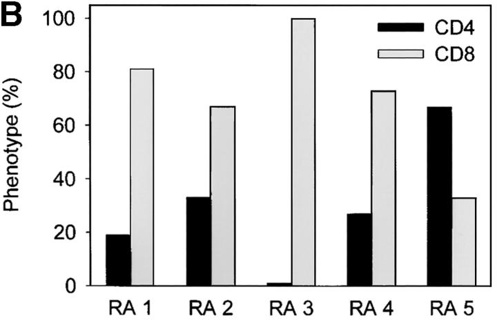
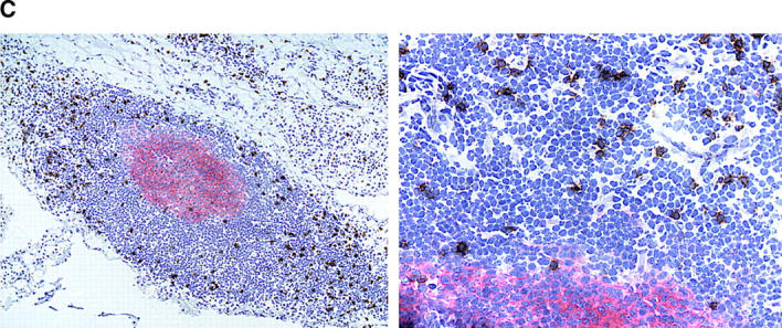
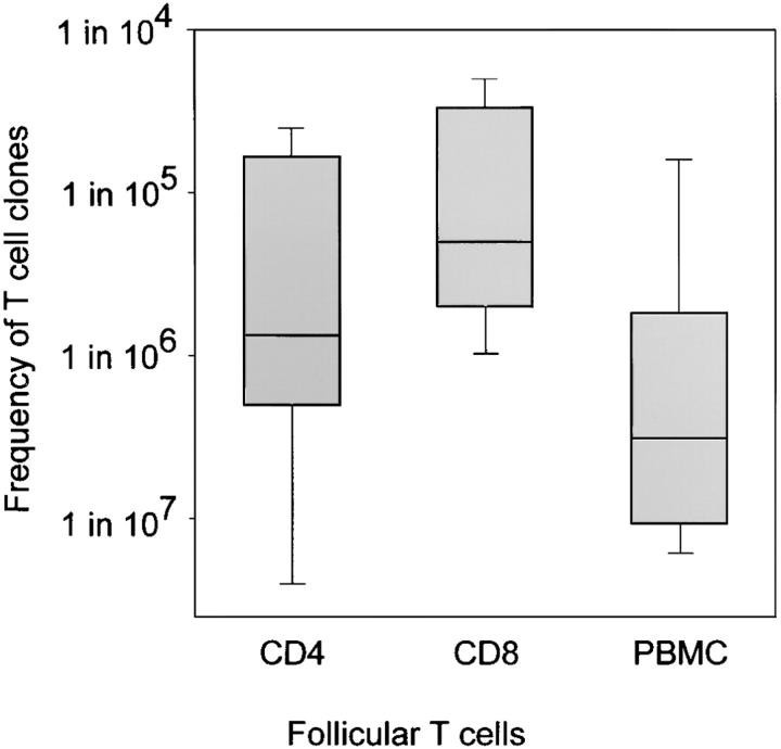
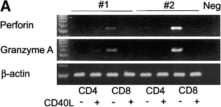
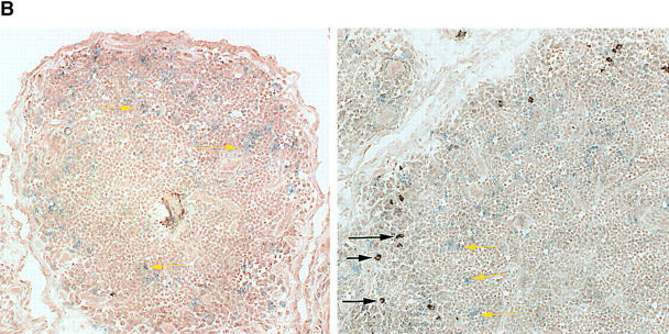
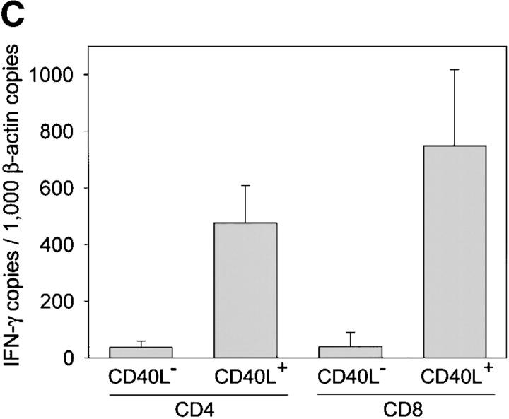
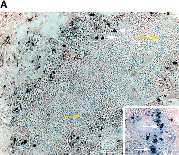
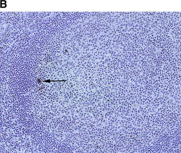
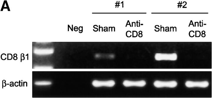
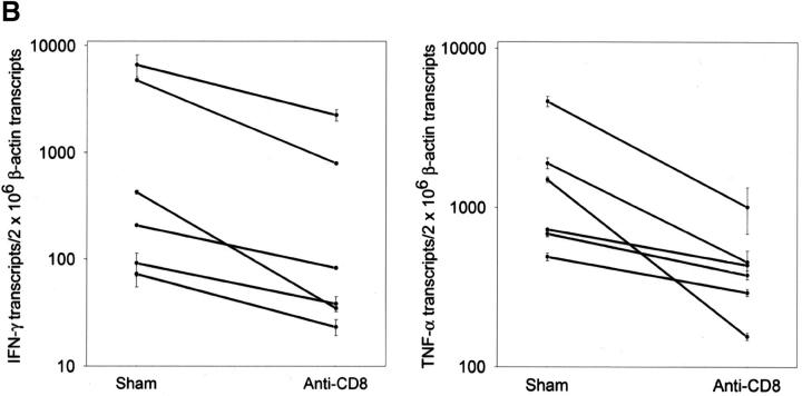
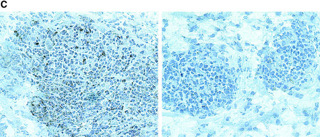
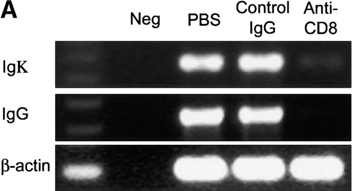
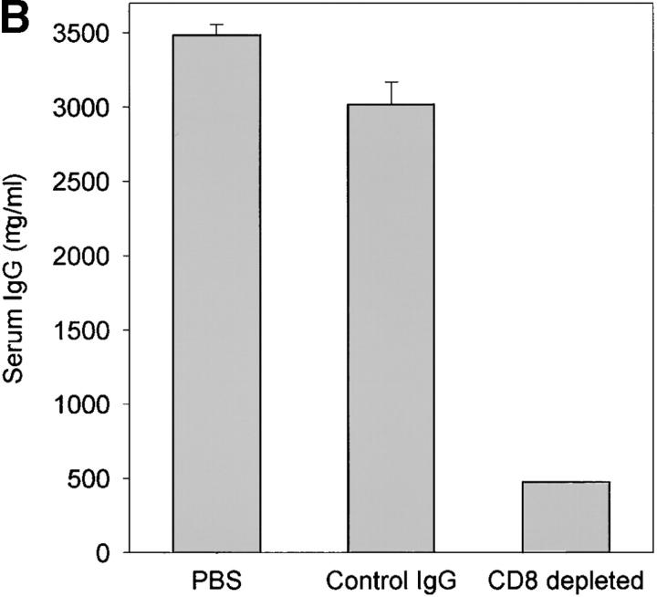
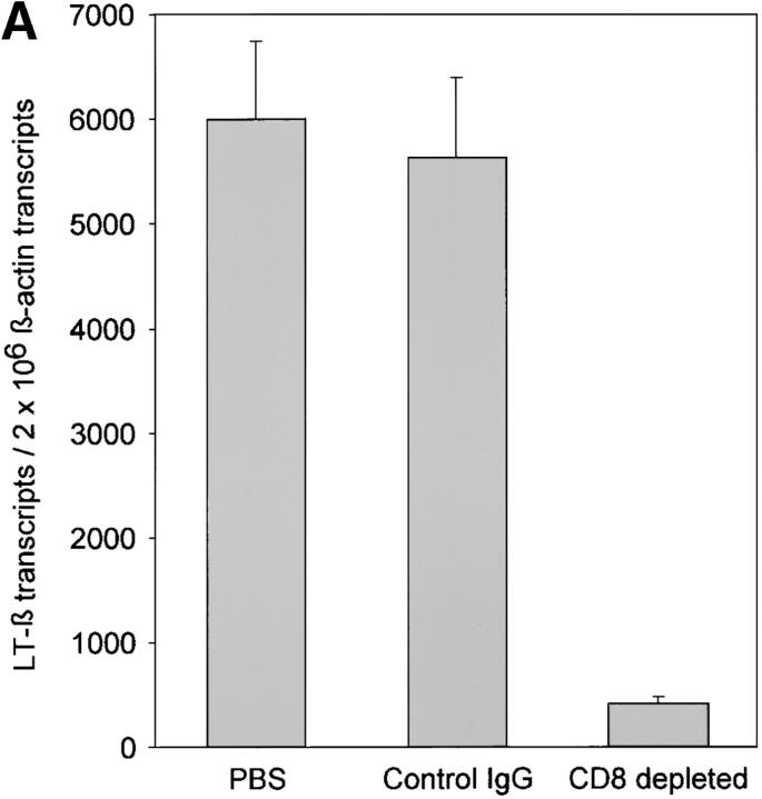
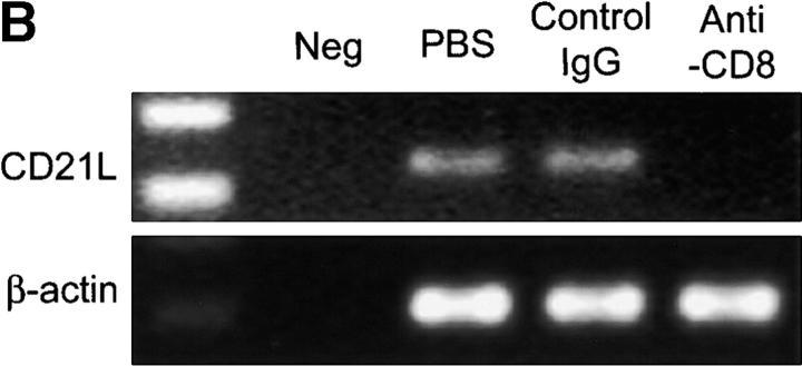
The assembly of inflammatory lesions in rheumatoid arthritis is highly regulated and typically leads to the formation of lymphoid follicles with germinal center (GC) reactions. We used microdissection of such extranodal follicles to analyze the colonizing T cells. Although the repertoire of follicular T cells was diverse, a subset of T cell receptor (TCR) sequences was detected in multiple independent follicles and not in interfollicular zones, suggesting recognition of a common antigen. Unexpectedly, the majority of shared TCR sequences were from CD8 T cells that were highly enriched in the synovium and present in low numbers in the periphery. To examine their role in extranodal GC reactions, CD8 T cells were depleted in human synovium-SCID mouse chimeras. Depletion of synovial CD8 T cells caused disintegration of the GC-containing follicles. In the absence of CD8 T cells, follicular dendritic cells disappeared, production of lymphotoxin-alpha1beta2 markedly decreased, and immunoglobulin (Ig) secretion ceased. Immunohistochemical studies demonstrated that these CD8 T cells accumulated at the edge of the mantle zone. Besides their unique localization, they were characterized by the production of interferon (IFN)-gamma, lack of the pore-forming enzyme perforin, and expression of CD40 ligand. Perifollicular IFN-gamma+ CD8 T cells were rare in secondary lymphoid tissues but accounted for the majority of IFN-gamma+ cells in synovial infiltrates. We propose that CD8+ T cells regulate the structural integrity and functional activity of GCs in ectopic lymphoid follicles.


Figures

Figure 1.
Figure 1.
Follicular TCR β-chain sequences preferentially derive from CD8 T cells. Synovial tissue was selected from five patients with RA who had multiple ectopic GCs in the synovial membrane. (A) The distribution of CD4 T cells (left), CD20 B cells (middle), and IgD+ cells (right) is shown in a representative example. The mantle zone is indicated by an asterisk (right). Follicles were microdissected and TCR β-chain sequences were obtained (Tables II and III). In parallel, synovial tissue and peripheral CD4 and CD8 cells were purified by FACS®. cDNA was tested for the presence of these TCR β-chain sequences by PCR-ELISA using N-D-N–specific oligonucleotides as probes. (B) In four of the five patients, the majority of the TCR β-chain sequences were detected in the CD8, and not the CD4, population. (C) To define the localization of the CD8 cells in synovial follicles, tissue sections were stained with anti-CD8 (brown) and anti-CD23 (red, expressed on follicular dendritic cells) mAb. CD8 T cells were found in the perifollicular zone, sometimes within the mantle zone, and only occasionally in the GC. Original magnifications: (left) × 100 and (right) × 400.
Figure 1.
Figure 1.
Follicular TCR β-chain sequences preferentially derive from CD8 T cells. Synovial tissue was selected from five patients with RA who had multiple ectopic GCs in the synovial membrane. (A) The distribution of CD4 T cells (left), CD20 B cells (middle), and IgD+ cells (right) is shown in a representative example. The mantle zone is indicated by an asterisk (right). Follicles were microdissected and TCR β-chain sequences were obtained (Tables II and III). In parallel, synovial tissue and peripheral CD4 and CD8 cells were purified by FACS®. cDNA was tested for the presence of these TCR β-chain sequences by PCR-ELISA using N-D-N–specific oligonucleotides as probes. (B) In four of the five patients, the majority of the TCR β-chain sequences were detected in the CD8, and not the CD4, population. (C) To define the localization of the CD8 cells in synovial follicles, tissue sections were stained with anti-CD8 (brown) and anti-CD23 (red, expressed on follicular dendritic cells) mAb. CD8 T cells were found in the perifollicular zone, sometimes within the mantle zone, and only occasionally in the GC. Original magnifications: (left) × 100 and (right) × 400.
Figure 2.
Figure 2.
Follicular T cells are relatively enriched in the synovial tissue. Frequencies of TCR β-chain sequences isolated from synovial follicles were determined in peripheral blood by limiting dilution. TCR amplification and hybridization with N-D-N–specific oligonucleotides was used to detect the presence of specific sequences. Results are shown as box plots displaying medians, 25th–75th percentile as the box and the 10th and 90th percentiles as whiskers. T cells expressing TCR β-chains derived from synovial tissue follicles were infrequent in peripheral blood. Sequences used by follicular CD8 T cells were slightly more frequent than those isolated from follicular CD4 T cells. Arbitrarily chosen TCR sequences from PBMCs of patients with RA have a median frequency of 1-in-5 × 10⁶ T cells. The enrichment of T cells with shared TCR β-chains in the synovial follicles is consistent with them being memory cells.
Figure 3.
Figure 3.
Functional profiles of synovial CD8+ T cell subsets. TCR sequences that were derived from synovial follicles were found in purified synovial CD8+CD40L+ T cells, suggesting that follicular CD8+ T cells may have a distinct functional profile (Table V). Synovial tissue CD8+ T cells were separated into CD40L+ and CD40L populations by FACS® and analyzed for the transcription of perforin, granzyme A, and IFN-γ. (A) Results for two patients are shown. Expression of perforin and granzyme A was restricted to CD8+CD40L T cells. (B) Immunohistochemistry confirmed that the majority of CD8 T cells (blue, yellow arrows) in the synovial lymphoid follicles lacked perforin expression. Perforin-positive cells (brown, black arrows) were absent in most follicles (left) and only occasionally seen in others (right). These perforin-positive cells did not coexpress CD8 and were presumably NK or NK T cells. Original magnification: × 200. (C) IFN-γ–specific transcripts were produced at significantly higher levels in CD40L+ than CD40L T cells. IFN-γ was semiquantified by PCR-ELISA and is shown as mean copy numbers ± SD of triplicate measurements.
Figure 3.
Figure 3.
Functional profiles of synovial CD8+ T cell subsets. TCR sequences that were derived from synovial follicles were found in purified synovial CD8+CD40L+ T cells, suggesting that follicular CD8+ T cells may have a distinct functional profile (Table V). Synovial tissue CD8+ T cells were separated into CD40L+ and CD40L populations by FACS® and analyzed for the transcription of perforin, granzyme A, and IFN-γ. (A) Results for two patients are shown. Expression of perforin and granzyme A was restricted to CD8+CD40L T cells. (B) Immunohistochemistry confirmed that the majority of CD8 T cells (blue, yellow arrows) in the synovial lymphoid follicles lacked perforin expression. Perforin-positive cells (brown, black arrows) were absent in most follicles (left) and only occasionally seen in others (right). These perforin-positive cells did not coexpress CD8 and were presumably NK or NK T cells. Original magnification: × 200. (C) IFN-γ–specific transcripts were produced at significantly higher levels in CD40L+ than CD40L T cells. IFN-γ was semiquantified by PCR-ELISA and is shown as mean copy numbers ± SD of triplicate measurements.
Figure 4.
Figure 4.
IFN-γ production by follicular CD8 T cells. (A) Synovial tissue sections were stained with anti-CD8 (blue) and anti–IFN-γ (brown) mAb. Most of the IFN-γ–producing cells expressed CD8 (white arrows). CD8+IFN-γ+ cells were located in the perifollicular zone at the outer edge of the mantle zone. (B) Yellow arrows mark CD8+IFN-γ cells. In contrast, IFN-γ–producing cells were extremely infrequent in tonsillar follicles, where they had a CD8 phenotype (black arrow). Original magnifications: × 200; inset, × 400.
Figure 4.
Figure 4.
IFN-γ production by follicular CD8 T cells. (A) Synovial tissue sections were stained with anti-CD8 (blue) and anti–IFN-γ (brown) mAb. Most of the IFN-γ–producing cells expressed CD8 (white arrows). CD8+IFN-γ+ cells were located in the perifollicular zone at the outer edge of the mantle zone. (B) Yellow arrows mark CD8+IFN-γ cells. In contrast, IFN-γ–producing cells were extremely infrequent in tonsillar follicles, where they had a CD8 phenotype (black arrow). Original magnifications: × 200; inset, × 400.
Figure 5.
Figure 5.
Depletion of synovial CD8 T cells suppresses IFN-γ and TNF-α production. Synovial tissues from patients with RA were engrafted into NOD-SCID mice. Chimeras were treated with anti-CD8 mAb; synovial tissue grafts were explanted after 7 d and analyzed for cytokine transcription. Anti-CD8 mAb treatment effectively depleted CD8 cells from the synovial tissue. (A) Transcripts for the CD8 β-chain were amplified by PCR in tissue extracts prepared from the grafts of sham or anti-CD8 treated chimeras. (B) After depletion of synovial CD8 T cells, in situ transcription of IFN-γ and TNF-α was significantly diminished. Results from six experiments with anti-CD8 mAb– and sham-treated mice are shown. Transcript numbers are adjusted relative to 2 × 10⁶ β-actin transcripts. Data are given as the mean ± SD of triplicate measurements by PCR-ELISA. (C) Immunohistochemical analysis of tissues retrieved from anti–CD8–treated mice (right) demonstrated that the tissues were depleted of IFN-γ+ cells (brown) in contrast to sham-treated mice (left). Ab-mediated depletion of CD8 T cells resulted in the disintegration of synovial follicles and the formation of cell clusters composed of dysmorphic lymphocytes.
Figure 5.
Figure 5.
Depletion of synovial CD8 T cells suppresses IFN-γ and TNF-α production. Synovial tissues from patients with RA were engrafted into NOD-SCID mice. Chimeras were treated with anti-CD8 mAb; synovial tissue grafts were explanted after 7 d and analyzed for cytokine transcription. Anti-CD8 mAb treatment effectively depleted CD8 cells from the synovial tissue. (A) Transcripts for the CD8 β-chain were amplified by PCR in tissue extracts prepared from the grafts of sham or anti-CD8 treated chimeras. (B) After depletion of synovial CD8 T cells, in situ transcription of IFN-γ and TNF-α was significantly diminished. Results from six experiments with anti-CD8 mAb– and sham-treated mice are shown. Transcript numbers are adjusted relative to 2 × 10⁶ β-actin transcripts. Data are given as the mean ± SD of triplicate measurements by PCR-ELISA. (C) Immunohistochemical analysis of tissues retrieved from anti–CD8–treated mice (right) demonstrated that the tissues were depleted of IFN-γ+ cells (brown) in contrast to sham-treated mice (left). Ab-mediated depletion of CD8 T cells resulted in the disintegration of synovial follicles and the formation of cell clusters composed of dysmorphic lymphocytes.
Figure 6.
Figure 6.
Ig production in synovial GCs is CD8 T cell dependent. Human synovium-SCID mouse chimeras were either sham-treated or treated with anti-CD8 mAb or isotype-matched control IgG as described in Fig. 5. Serum and synovial tissues were collected from the mice 7 d after the treatment.(A) cDNA from the tissues were amplified with Igκ- and IgG-specific primer sets in multiplex PCR. Anti-CD8 mAb treatment but not treatment with control IgG clearly diminished the transcription of Ig H and L chains. (B) Human IgG was quantified in serum from the treated and sham-treated mice by ELISA. Tissue depleted of CD8 T cells secreted minimal amounts of human IgG. Results are shown as mean ± SD of triplicate measurements. Similar results were seen in experiments with tissues from two additional patients.
Figure 6.
Figure 6.
Ig production in synovial GCs is CD8 T cell dependent. Human synovium-SCID mouse chimeras were either sham-treated or treated with anti-CD8 mAb or isotype-matched control IgG as described in Fig. 5. Serum and synovial tissues were collected from the mice 7 d after the treatment.(A) cDNA from the tissues were amplified with Igκ- and IgG-specific primer sets in multiplex PCR. Anti-CD8 mAb treatment but not treatment with control IgG clearly diminished the transcription of Ig H and L chains. (B) Human IgG was quantified in serum from the treated and sham-treated mice by ELISA. Tissue depleted of CD8 T cells secreted minimal amounts of human IgG. Results are shown as mean ± SD of triplicate measurements. Similar results were seen in experiments with tissues from two additional patients.
Figure 7.
Figure 7.
Depletion of CD8 T cells disrupts synovial GCs. GCs depend on the production of LT-α1β2 and the presence of FDCs. LT-β transcripts were semiquantified by PCR-ELISA. (A) Treatment with anti-CD8 mAb significantly reduced the relative number of LT-β transcripts in the tissues. Results from one of three experiments are shown as mean ± SD of triplicate measurements. CD8 T cell depletion abrogated CD21L transcription, confirming that FDC networks were no longer present. (B) Synovial tissues retrieved from control and anti-CD8–treated chimeras were assayed for the presence of CD21L transcript, which is exclusively produced by FDCs.
Figure 7.
Figure 7.
Depletion of CD8 T cells disrupts synovial GCs. GCs depend on the production of LT-α1β2 and the presence of FDCs. LT-β transcripts were semiquantified by PCR-ELISA. (A) Treatment with anti-CD8 mAb significantly reduced the relative number of LT-β transcripts in the tissues. Results from one of three experiments are shown as mean ± SD of triplicate measurements. CD8 T cell depletion abrogated CD21L transcription, confirming that FDC networks were no longer present. (B) Synovial tissues retrieved from control and anti-CD8–treated chimeras were assayed for the presence of CD21L transcript, which is exclusively produced by FDCs.

Similar articles

Cited by

References

    1. Nepom, G.T. 1998. Major histocompatibility complex-directed susceptibility to rheumatoid arthritis. Adv. Immunol. 68:315–332. - PubMed
    1. Weyand, C.M., T.G. McCarthy, and J.J. Goronzy. 1995. Correlation between disease phenotype and genetic heterogeneity in rheumatoid arthritis. J. Clin. Invest. 95:2120–2126. - PMC - PubMed
    1. Yen, J.H., B.E. Moore, T. Nakajima, D. Scholl, D.J. Schaid, C.M. Weyand, and J.J. Goronzy. 2001. Major histocompatibility complex class I–recognizing receptors are disease risk genes in rheumatoid arthritis. J. Exp. Med. 193:1159–1167. - PMC - PubMed
    1. Young, C.L., T.C. Adamson, J.H. Vaughan, and R.I. Fox. 1984. Immunohistologic characterization of synovial membrane lymphocytes in rheumatoid arthritis. Arthr. Rheum. 27:32–39. - PubMed
    1. Schroder, A.E., A. Greiner, C. Seyfert, and C. Berek. 1996. Differentiation of B cells in the nonlymphoid tissue of the synovial membrane of patients with rheumatoid arthritis. Proc. Natl. Acad. Sci. USA. 93:221–225. - PMC - PubMed

Publication types

MeSH terms

Substances