. 2002 May 20;195(10):1359-70.
doi: 10.1084/jem.20010753.

Modulation of human immunodeficiency virus 1 replication by interferon regulatory factors

Affiliations

Modulation of human immunodeficiency virus 1 replication by interferon regulatory factors

Marco Sgarbanti et al. J Exp Med. .

Abstract

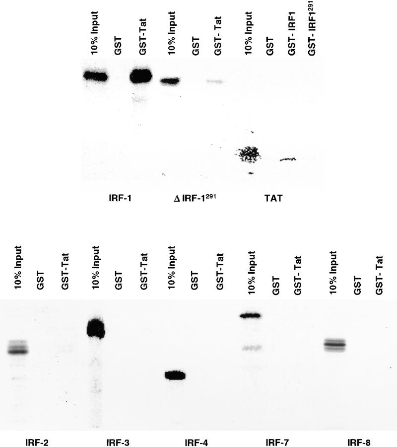
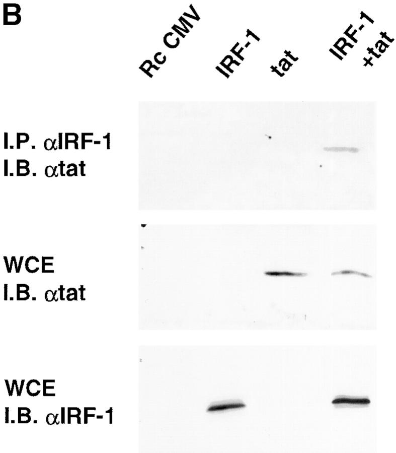
Transcription of the human immunodeficiency virus (HIV)-1 is controlled by the cooperation of virally encoded and host regulatory proteins. The Tat protein is essential for viral replication, however, expression of Tat after virus entry requires HIV-1 promoter activation. A sequence in the 5' HIV-1 LTR, containing a binding site for transcription factors of the interferon regulatory factors (IRF) family has been suggested to be critical for HIV-1 transcription and replication. Here we show that IRF-1 activates HIV-1 LTR transcription in a dose-dependent fashion and in the absence of Tat. This has biological significance since IRF-1 is produced early upon virus entry, both in cell lines and in primary CD4+ T cells, and before expression of Tat. IRF-1 also cooperates with Tat in amplifying virus gene transcription and replication. This cooperation depends upon a physical interaction that is blocked by overexpression of IRF-8, the natural repressor of IRF-1, and, in turn is released by overexpression of IRF-1. These data suggest a key role of IRF-1 in the early phase of viral replication and/or during viral reactivation from latency, when viral transactivators are absent or present at very low levels, and suggest that the interplay between IRF-1 and IRF-8 may play a key role in virus latency.


Figures

Figure 1.
Figure 1.
Effect of IRFs on HIV-1 LTR transactivation. (A) Jurkat cells were transiently cotransfected with the HIV-1 LTR-CAT (1 μg) and vectors (2 μg) expressing the indicated IRFs. IRF-3 5D and IRF-7* codify for the constitutively activated forms of IRF-3 and -7, respectively (references and 41). After 24 h, CAT activity was evaluated as indicated in the Materials and Methods. (B) Dose–response effect of the wild-type IRF-1 or its mutant deleted in the activation domain (Δ IRF-1) on HIV-1 LTR-directed gene expression. Cells were transfected as in A except that 0.8 μg of HIV-1 LTR-CAT and increasing amounts of the vectors expressing IRF-1 or Δ IRF-1 (0.8, 1.6, 3.2 μg) were used. (C) Effect of IRF-1 on mutated HIV-LTR constructs. Cells were transiently transfected with wild-type HIV-LTR or Δ1 LTR in which the ISRE region is deleted (reference 9), Δ2 LTR in which the NF-κB sites are mutated or Δ3 LTR in which both sites are deleted/mutated. IRF-1–expressing vector was cotransfected as indicated. Results are shown as percentages of the CAT activity of the wild-type HIV-LTR in IRF-1–transfected cells. (D) IRF-1 cooperates with Tat to enhance HIV-1 LTR-CAT activity. Cells were transiently cotransfected with the HIV-1 LTR-CAT (1 μg), Tat (5 ng), or IRF-1 (1 μg) expressing vectors, either alone or in combination, as indicated. CAT activity was determined after 24 h. The results quantified by an Instant Imager are reported as mean levels ± SE from three separate experiments.
Figure 1.
Figure 1.
Effect of IRFs on HIV-1 LTR transactivation. (A) Jurkat cells were transiently cotransfected with the HIV-1 LTR-CAT (1 μg) and vectors (2 μg) expressing the indicated IRFs. IRF-3 5D and IRF-7* codify for the constitutively activated forms of IRF-3 and -7, respectively (references and 41). After 24 h, CAT activity was evaluated as indicated in the Materials and Methods. (B) Dose–response effect of the wild-type IRF-1 or its mutant deleted in the activation domain (Δ IRF-1) on HIV-1 LTR-directed gene expression. Cells were transfected as in A except that 0.8 μg of HIV-1 LTR-CAT and increasing amounts of the vectors expressing IRF-1 or Δ IRF-1 (0.8, 1.6, 3.2 μg) were used. (C) Effect of IRF-1 on mutated HIV-LTR constructs. Cells were transiently transfected with wild-type HIV-LTR or Δ1 LTR in which the ISRE region is deleted (reference 9), Δ2 LTR in which the NF-κB sites are mutated or Δ3 LTR in which both sites are deleted/mutated. IRF-1–expressing vector was cotransfected as indicated. Results are shown as percentages of the CAT activity of the wild-type HIV-LTR in IRF-1–transfected cells. (D) IRF-1 cooperates with Tat to enhance HIV-1 LTR-CAT activity. Cells were transiently cotransfected with the HIV-1 LTR-CAT (1 μg), Tat (5 ng), or IRF-1 (1 μg) expressing vectors, either alone or in combination, as indicated. CAT activity was determined after 24 h. The results quantified by an Instant Imager are reported as mean levels ± SE from three separate experiments.
Figure 2.
Figure 2.
IRF-1 mRNA is induced early upon HIV-1 infection and before expression of Tat. (A) Jurkat cells were infected with the HIV-1 strain IIIB (5,000 cpm/ml) and, at the indicated time points, total RNA was extracted and analyzed by RNase protection with a IRF-1–specific antisense riboprobe. 18S rRNA was used as a control of RNA loading. (B) mRNA relative fold-increase after normalization to the 18S RNA, quantified by Instant Imager. Mean values from three separate experiments are shown. (C) Total RNA extracted at 5 and 24 h after infection shown in A was analyzed by RT-PCR for the doubly-spliced (tat/rev) transcript as described in Materials and Methods.
Figure 2.
Figure 2.
IRF-1 mRNA is induced early upon HIV-1 infection and before expression of Tat. (A) Jurkat cells were infected with the HIV-1 strain IIIB (5,000 cpm/ml) and, at the indicated time points, total RNA was extracted and analyzed by RNase protection with a IRF-1–specific antisense riboprobe. 18S rRNA was used as a control of RNA loading. (B) mRNA relative fold-increase after normalization to the 18S RNA, quantified by Instant Imager. Mean values from three separate experiments are shown. (C) Total RNA extracted at 5 and 24 h after infection shown in A was analyzed by RT-PCR for the doubly-spliced (tat/rev) transcript as described in Materials and Methods.
Figure 4.
Figure 4.
IRF-1 and IRF-2 bind the HIV-ISRE. (A) Nuclear cell extracts (20 μg) from Jurkat cells uninfected or infected with HIV-1 IIIB strain (5,000 cpm/ml) were prepared at different time points after infection and incubated with an oligonucleotide corresponding to the four tandem IRF-binding sites (C13). Supershift assays were performed in the presence of specific anti–IRF-1 and anti–IRF-2 antibodies as indicated. Binding complexes were resolved by PAGE and visualized by autoradiography. (B) DNA pull-down assays. Biotinylated oligodeoxynucleotides containing the wild-type or a mutated version of the HIV-ISRE (Materials and Methods), coupled to Streptavidin MagneSphere were incubated with nuclear extracts from Jurkat cells or primary CD4+ T cells infected with the HIV-1 IIIB strain (5,000 cpm/ml). The bound proteins were eluted from the beads by boiling in sample buffer and analyzed by WB with antibodies against IRF-1. INPUT indicates the level of endogenous IRF-1 in the uninfected and infected nuclear cell extracts (20 μg) at the indicated time points determined by WB analysis.
Figure 3.
Figure 3.
HIV-1 infection induces IRF-1 mRNA expression in primary CD4+ T lymphocytes. (A) Purified CD4+ T cells were infected with the HIV-1 IIIB strain (5,000 cpm/ml) and IRF-1 mRNA evaluated by semiquantitative RT-PCR analysis at the indicated time points after infection. A representative experiment out of three performed is shown. The increase in IRF-1 mRNA accumulation begun to be evident between 5 and 12 h after infection and peaked between 24 and 48 h after infection depending on the donor. 26S RNA was used for normalization as described in Materials and Methods. (B) mRNA relative fold-increase after normalization to the 26S RNA quantified by Instant Imager. (C) Total RNA extracted at the indicated time points as in A was analyzed by RT-PCR for the doubly-spliced (tat/rev) transcript as described in Materials and Methods.
Figure 3.
Figure 3.
HIV-1 infection induces IRF-1 mRNA expression in primary CD4+ T lymphocytes. (A) Purified CD4+ T cells were infected with the HIV-1 IIIB strain (5,000 cpm/ml) and IRF-1 mRNA evaluated by semiquantitative RT-PCR analysis at the indicated time points after infection. A representative experiment out of three performed is shown. The increase in IRF-1 mRNA accumulation begun to be evident between 5 and 12 h after infection and peaked between 24 and 48 h after infection depending on the donor. 26S RNA was used for normalization as described in Materials and Methods. (B) mRNA relative fold-increase after normalization to the 26S RNA quantified by Instant Imager. (C) Total RNA extracted at the indicated time points as in A was analyzed by RT-PCR for the doubly-spliced (tat/rev) transcript as described in Materials and Methods.
Figure 5.
Figure 5.
GST-pull down assays. The indicated IRFs (IRF-1, IRF-2, IRF-3, IRF-4, IRF-7, IRF-8, a COOH-terminal deleted mutant of IRF-1 [Δ IRF-1]), and Tat, were translated in vitro in the presence of 35[S] methionine as indicated in Materials and Methods and incubated with recombinant GST–Tat or GST-IRF-1 fusion proteins immobilized on glutathione-sepharose. Input corresponds to 10% of the 35[S]-labeled proteins used in the binding experiments. The complexes were resolved by PAGE and detected by autoradiography. Binding of 35[S]-labeled proteins to beads containing only GST protein is also shown. Quantitation of incorporated radioactivity was performed by Instant Imager.
Figure 6.
Figure 6.
IRF-1 and Tat associate intracellularly. (A) In vitro–translated IRF-1 (lanes 1–3) and nuclear cell extracts from Jurkat cells treated with IFN-γ (lanes 4–6) or control medium (lanes 7–9) were incubated with purified GST–Tat fusion protein or GST alone. Bound proteins were then analyzed by WB using anti–IRF-1 polyclonal antibody as described in Materials and Methods. 10% of the extract used for binding assays is shown in lane 7 (untreated cells) and lane 4 (IFN-γ-treated cells). The slowly migrating IRF-1p* band observed in IFN-γ–treated cell extracts is due to the phosphorylation induced by IFN-γ (reference 64). Extra bands in lanes 7–9 are not specific. (B) 293 HEK cells were transfected with the expression plasmids encoding IRF-1 or Tat, alone or in combination. Whole cell extracts (300 μg) were immunoprecipitated with anti–IRF-1 antibodies (αIRF-1). Immunoprecipitated complexes were separated by 10% SDS-PAGE and subsequently probed with anti-Tat antibodies (αTat) as indicated. Whole cell extracts (10 μg) were separated on 10% or 15% SDS-PAGE and probed with anti-Tat or anti–IRF-1 antibodies.
Figure 6.
Figure 6.
IRF-1 and Tat associate intracellularly. (A) In vitro–translated IRF-1 (lanes 1–3) and nuclear cell extracts from Jurkat cells treated with IFN-γ (lanes 4–6) or control medium (lanes 7–9) were incubated with purified GST–Tat fusion protein or GST alone. Bound proteins were then analyzed by WB using anti–IRF-1 polyclonal antibody as described in Materials and Methods. 10% of the extract used for binding assays is shown in lane 7 (untreated cells) and lane 4 (IFN-γ-treated cells). The slowly migrating IRF-1p* band observed in IFN-γ–treated cell extracts is due to the phosphorylation induced by IFN-γ (reference 64). Extra bands in lanes 7–9 are not specific. (B) 293 HEK cells were transfected with the expression plasmids encoding IRF-1 or Tat, alone or in combination. Whole cell extracts (300 μg) were immunoprecipitated with anti–IRF-1 antibodies (αIRF-1). Immunoprecipitated complexes were separated by 10% SDS-PAGE and subsequently probed with anti-Tat antibodies (αTat) as indicated. Whole cell extracts (10 μg) were separated on 10% or 15% SDS-PAGE and probed with anti-Tat or anti–IRF-1 antibodies.
Figure 7.
Figure 7.
IRF-8 but not IRF-2 inhibits the IRF-1-mediated and Tat-mediated HIV-1 LTR activity. (A and B) Transient cotransfections were performed with the HIV-LTR CAT reporter construct (1 μg) and IRF-1, IRF-2, IRF-8 (1 μg), or Tat (5 ng) expression vectors, respectively, as indicated. CAT activity was quantified 48 h after transfection. (C) RNase protection assay with a IRF-8–specific antisense riboprobe on total RNA extracted from Jurkat cells transfected with an empty vector or an IRF-8–expressing vector. IRF-8 indicates the transcript of the transduced gene and IRF-8e the endogenous recognized transcript. 18S RNA was used as a control of RNA loading and tRNA as a control of specificity. (D) Jurkat cells constitutively expressing IRF-8 or the empty vector were transiently transfected with the Tat-expressing vector (20 ng) or IRF-1–expressing vector (1 μg) along with the HIV-LTR reporter construct and CAT activity quantified as described in B. The results quantified by an Instant Imager are reported as mean levels ± SE from three separate experiments.
Figure 7.
Figure 7.
IRF-8 but not IRF-2 inhibits the IRF-1-mediated and Tat-mediated HIV-1 LTR activity. (A and B) Transient cotransfections were performed with the HIV-LTR CAT reporter construct (1 μg) and IRF-1, IRF-2, IRF-8 (1 μg), or Tat (5 ng) expression vectors, respectively, as indicated. CAT activity was quantified 48 h after transfection. (C) RNase protection assay with a IRF-8–specific antisense riboprobe on total RNA extracted from Jurkat cells transfected with an empty vector or an IRF-8–expressing vector. IRF-8 indicates the transcript of the transduced gene and IRF-8e the endogenous recognized transcript. 18S RNA was used as a control of RNA loading and tRNA as a control of specificity. (D) Jurkat cells constitutively expressing IRF-8 or the empty vector were transiently transfected with the Tat-expressing vector (20 ng) or IRF-1–expressing vector (1 μg) along with the HIV-LTR reporter construct and CAT activity quantified as described in B. The results quantified by an Instant Imager are reported as mean levels ± SE from three separate experiments.
Figure 8.
Figure 8.
Inhibition of HIV-1 replication in IRF-8–expressing Jurkat cells. Jurkat cells stably transfected with the IRF-8 (lanes 2, 4, and 6) or the RcCMV (control vector) (lanes 1, 3, and 5) were infected with the HIV-1 IIIB strain at an infectious dose corresponding to 1,000 or 5,000 cpm/ml of RT activity. (A) Cells were collected after 24 and 48 h and total RNA analyzed by RT-PCR, as described in Materials and Methods. (B) HIV-p24 antigen production. After 48, 72 and 144 h, p24 antigen accumulation was determined in the cell supernatants as indicated in Materials and Methods.
Figure 8.
Figure 8.
Inhibition of HIV-1 replication in IRF-8–expressing Jurkat cells. Jurkat cells stably transfected with the IRF-8 (lanes 2, 4, and 6) or the RcCMV (control vector) (lanes 1, 3, and 5) were infected with the HIV-1 IIIB strain at an infectious dose corresponding to 1,000 or 5,000 cpm/ml of RT activity. (A) Cells were collected after 24 and 48 h and total RNA analyzed by RT-PCR, as described in Materials and Methods. (B) HIV-p24 antigen production. After 48, 72 and 144 h, p24 antigen accumulation was determined in the cell supernatants as indicated in Materials and Methods.
Figure 9.
Figure 9.
IRF-8 inhibits the binding of IRF-1 to immobilized Tat. Recombinant GST–Tat fusion protein was immobilized on glutathione agarose beads and incubated with the indicated 35[S]-labeled IRFs as described in Materials and Methods. Input corresponds to 10% of the 35[S]-labeled proteins used in the binding experiments; in lanes 1, 5, and 6 are shown the in vitro labeled IRF-8, IRF-2, and IRF-1, respectively. In lane 2, 35[S]-labeled IRF-1 and IRF-8 were incubated together for 15 min at room temperature before the addition of GST–Tat fusion protein beads. Lane 3 shows the binding of 35[S]-labeled IRF-1 alone to GST–Tat beads. In lane 4, 35[S]-labeled IRF-1 and IRF-2 were preincubated together for 15 min at room temperature before the addition of GST–Tat fusion protein beads.
Figure 10.
Figure 10.
IRF-1 overexpression releases the inhibition of HIV-1 replication by IRF-8. Bulk populations of Jurkat cells stably transfected with IRF-8 were engineered to constitutively express IRF-1. (A) RNase protection with a IRF-1–specific antisense riboprobe on total RNA extracted from IRF-8–expressing cells, transfected with an empty vector (RcIRF-8/Rc), or with an IRF-1–expressing vector (RcIRF-8/IRF-1). 18S RNA was used as a control of RNA loading and tRNA as a control of specificity. (B) Cells were infected with the HIV-1 IIIB strain as in Fig. 8 and after 48, 72, and 144 h, HIV p24 antigen accumulation was determined in the cell supernatants as indicated in Materials and Methods.
Figure 10.
Figure 10.
IRF-1 overexpression releases the inhibition of HIV-1 replication by IRF-8. Bulk populations of Jurkat cells stably transfected with IRF-8 were engineered to constitutively express IRF-1. (A) RNase protection with a IRF-1–specific antisense riboprobe on total RNA extracted from IRF-8–expressing cells, transfected with an empty vector (RcIRF-8/Rc), or with an IRF-1–expressing vector (RcIRF-8/IRF-1). 18S RNA was used as a control of RNA loading and tRNA as a control of specificity. (B) Cells were infected with the HIV-1 IIIB strain as in Fig. 8 and after 48, 72, and 144 h, HIV p24 antigen accumulation was determined in the cell supernatants as indicated in Materials and Methods.

Similar articles

Cited by

References

    1. Cullen, B.R. 1991. Regulation of HIV-1 gene expression. FASEB J. 5:2361–2368. - PubMed
    1. Van Lint, C., J. Ghysdael, P. Paras, Jr., A. Burnyand, and E. Verdin. 1994. A transcriptional regulatory element is associated with a nuclease-hypersensitive site in the pol gene of human immunodeficiency virus type. J. Virol. 68:2632–2648. - PMC - PubMed
    1. Jones, K.A., and B.M. Peterlin. 1994. Control of RNA initiation and elongation at the HIV-1 promoter. Annu. Rev. Biochem. 63:717–743. - PubMed
    1. Gaynor, R. 1992. Cellular transcription factors involved in the regulation of HIV-1 gene expression. AIDS. 6:347–363. - PubMed
    1. el Kharroubi, A., and E. Verdin. 1994. Protein-DNA interactions within DNase I-hypersensitive sites located downstream of the HIV-1 promoter. J. Biol. Chem. 269:19916–19924. - PubMed

Publication types

MeSH terms