. 2002 Jun;76(12):5875-81.
doi: 10.1128/jvi.76.12.5875-5881.2002.

Regulation of hepatocyte nuclear factor 1 activity by wild-type and mutant hepatitis B virus X proteins

Affiliations

Regulation of hepatocyte nuclear factor 1 activity by wild-type and mutant hepatitis B virus X proteins

Jie Li et al. J Virol. 2002 Jun.

Abstract

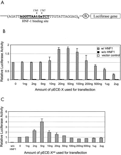
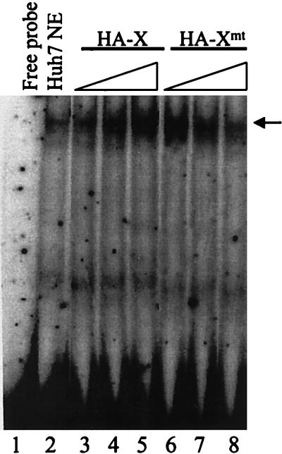
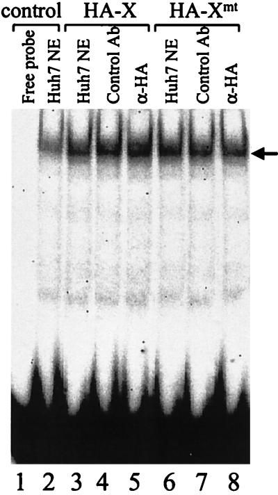
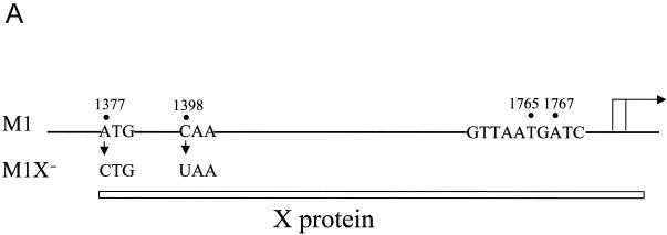
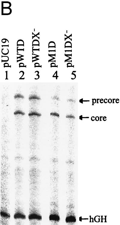
The hepatitis B virus (HBV) core promoter regulates the transcription of two related RNA products named precore RNA and core RNA. Previous studies indicate that a double-nucleotide mutation that occurs frequently during chronic HBV infection converts a nuclear receptor binding site in the core promoter to the binding site of the transcription factor hepatocyte nuclear factor-1 (HNF-1) and specifically suppresses the transcription of the precore RNA. This mutation also changes two codons in the overlapping X protein coding sequence. In this report, we demonstrate that the X protein and its mutant X(mt) can physically bind to HNF-1 both in vitro and in vivo. Further analyses indicate that both X and X(mt) can enhance the gene transactivation and the DNA binding activities of HNF-1. This finding demonstrates for the first time that the X protein can stimulate the DNA binding activity of a homeodomain transcription factor. Interestingly, while both X and X(mt) can stimulate the HNF-1 activities, they differ in their effects: a smaller amount of X(mt) is needed to generate greater transactivation and DNA binding activities of HNF-1. This functional difference between X and X(mt) may have important implications in HBV pathogenesis and is apparently why they have different effects on the core promoter bearing the HNF-1 binding site.


Figures

FIG. 1.
FIG. 1.
Coimmunoprecipitation of HNF-1 with X and Xmt proteins in vitro. HNF-1, HA-X, and HA-Xmt were labeled with [35S]methionine and synthesized in vitro with rabbit reticulocyte lysates. Details of the procedures are described in Materials and Methods. They were then subjected to gel electrophoresis without immunoprecipitation (lanes 1 to 3) or after immunoprecipitation (lanes 4 to 8) with the anti-HA antibody. Lane 1, HNF-1 without immunoprecipitation; lane 2, HA-X without immunoprecipitation; lane 3, HA-Xmt without immunoprecipitation; lane 4, HNF-1 with immunoprecipitation with the anti-HA antibody; lane 5, HA-X with immunoprecipitation; lane 6, HA-Xmt with immunoprecipitation; lane 7, HA-X and HNF-1 immunoprecipitated with the anti-HA antibody; and lane 8, HA-Xmt and HNF-1 immunoprecipitated with the anti-HA antibody. The arrows mark the locations of the HNF-1, HA-X, and HA-Xmt bands. In lane 1, protein bands smaller than the size of HNF-1 were also detected. The nature of these bands is unclear. They might be degraded HNF-1 or N-terminally truncated HNF-1 synthesized from the internal ATG codons. Sizes are shown on the left in kilodaltons.
FIG. 2.
FIG. 2.
Coimmunoprecipitation of HNF-1 with X and Xmt expressed in Huh7 cells. (A) Coimmunoprecipitation with anti-HA antibody as the primary antibody. Huh7 cells were transfected with the control pRc/CMV vector alone (lane 1), pCMV-HNF-1 (lanes 2 and 7), pCMV-HAX (lane 3), pCMV-HAXmt (lane 4), pCMV-HNF-1 and pCMV-HAX (lane 5), or pCMV-HNF-1 and pCMV-HAXmt (lane 6). Two days after transfection, cells were lysed, immunoprecipitated with the mouse anti-HA antibody, and then analyzed by Western blotting with the rabbit anti-HNF-1 antibody (lanes 1 to 6). In lane 7, the lysates of cells transfected by pCMV-HNF-1 were analyzed directly by Western blotting without immunoprecipitation. This lane served as a positive control for identifying the HNF-1 protein band. The arrow marks the location of a background band. (B) Coimmunoprecipitation with the anti-HNF-1 antibody as the primary antibody. The coimmunoprecipitation procedures were the same as in panel A except that the rabbit anti-HNF-1 antibody was used for immunoprecipitation, followed by Western blotting with the anti-HA antibody. Lane 1, HA-tagged X protein purified from E. coli, which served as a marker; lane 2, Huh7 cells transfected with pRc/CMV control vector; lane 3, Huh7 cells transfected with pCMV-HNF-1; lane 4, Huh7 cells transfected with pCMV-HNF-1 and pCMV-HAX; lane 5, cells transfected with pCMV-HNF-1 and pCMV-HAXmt; lane 6, cell lysates prepared from cells transfected with pCMV-HAX without immunoprecipitation with the anti-HNF-1 antibody.
FIG. 3.
FIG. 3.
Effects of X and Xmt on the transactivation activities of HNF-1. (A) Illustration of the HNF-1 reporter construct. The luciferase reporter is boxed, and the HNF-1 binding sequence is shown in boldface letters and underlined. A minimal thymidine kinase (tk) promoter controlled the expression of the luciferase reporter. (B) Histogram of the transactivation activities of X and HNF-1. Shaded box, cotransfection of pCpHNF1-Luc and pCMV-HNF-1; solid box, transfection of pCpHNF1-Luc with pRc/CMV; empty box, transfection of the control reporter lacking the HNF-1 binding site. (C) Histogram of the transactivation activities of Xmt and HNF-1. In both B and C, 3 μg of pCMV-HNF-1 or pRc/CMV was used for the cotransfection with 4 μg of pCpHNF1-Luc or the control reporter ptk-Luc. The amount of pECE-X or pECE-Xmt used for the transfection is indicated at the bottom of the chart. In all cases, 40 ng of pXGH5, a plasmid that expressed the human growth hormone, was also used for cotransfection to monitor the transfection efficiency. All the luciferase activities expressed were normalized against the luciferase activity derived from the cotransfection of pCMV-HNF-1, pCpHNF1-Luc, and pECE-1. The results represent the averages of at least three independent experiments.
FIG. 4.
FIG. 4.
HA-X and HA-Xmt proteins expressed in E. coli. The procedures for the expression of HA-X and HA-Xmt in E. coli and their subsequent purifications are described in Materials and Methods. Lanes 1 and 2, silver staining of HA-X (lane 1) and HA-Xmt (lane 2) purified from E. coli. Lanes 3 and 4, Western blot analysis of HA-X (lane 3) and HA-Xmt (lane 4) with the anti-HA antibody. The arrow marks the locations of HA-X and HA-Xmt. Sizes are shown in kilodaltons.
FIG. 5.
FIG. 5.
Enhancement of the DNA binding activity of HNF-1 by HA-X and HA-Xmt. The procedures for the gel shift experiment are described in Materials and Methods. Lane 1, free DNA probe; lane 2, free DNA probe mixed with the Huh7 nuclear extracts; lanes 3 to 5, the addition of 0.2 ng, 20 ng, and 100 ng of HA-X, respectively, into the binding reaction mixture; lanes 6 to 8, the addition of 0.2 ng, 20 ng, and 100 ng of HA-Xmt, respectively, into the binding reaction mixture. The location of the HNF-1 band shift is indicated by an arrow.
FIG. 6.
FIG. 6.
Absence of HA-X and HA-Xmt in the HNF-1 band shift. The supershift assay was conducted as described in Materials and Methods. Lane 1, free DNA probe; lane 2, DNA probe incubated with Huh7 nuclear extract (NE); lane 3, Huh7 nuclear extract plus 1 ng of X protein; lane 4, the same as lane 3 but with the addition of a control mouse antibody; lane 5, the same as lane 4 but with the addition of the anti-HA antibody; lane 6, nuclear extracts plus 1 ng of HA-Xmt protein; lane 7, the same as lane 6 but with the addition of a control mouse antibody; lanes 8, the same as lane 7 but with the addition of the anti-HA antibody. The location of the HNF-1 band shifts is indicated by an arrow.
FIG. 7.
FIG. 7.
Effects of X and Xmt on transcription of precore and core RNAs. (A) Illustration of the HBV genome with the mutations that abolished the expression of the Xmt protein. The locations of the nt 1376 A to C and nt 1397 C to U double mutations that abolished the expression of the Xmt protein are indicated. The HNF-1 binding sequence in the M1 genome is also shown. The rightward arrow marks the locations of precore RNA and core RNA transcription initiation sites. (B) Primer extension analysis of precore RNA and core RNA transcribed from various HBV genomic DNA constructs. Huh7 cells transfected with various HBV genomic dimers were lysed 2 days after transfection. The total cellular RNA was then extracted and used for the primer extension analysis. Lane 1, cells transfected with the control pUC19 vector; lane 2, cells transfected with the wild-type HBV genome (pWTD); lane 3, cells transfected with the wild-type HBV genome with mutations that abolished the expression of the X protein (pWTDX); lane 4, the M1 HBV genome (pM1D); lane 5, the M1 genome with mutations that abolished Xmt protein expression (pM1DX). In all cases, pXGH5, a plasmid that expresses the human growth hormone (hGH), was used for cotransfection to serve as the internal transfection control. The locations of the precore RNA and the core RNA as well as the internal control human growth hormone RNA are indicated by arrows. The procedures for the primer extension analysis are described in Materials and Methods. (C) Quantitation analysis of the results shown in panel B. The signals of the precore (PC) RNA and the core (C) RNA bands were measured by densitometry and calculated based on the equation [(XaB)/(XWTDB)]/[TEa/TEWTD], where Xa is the precore RNA (or the core RNA) signal of sample a, XWTD is the precore RNA (or the core RNA) signal expressed by pWTD, B is the background signal from a randomly selected area of the gel, TEa is the transfection efficiency of sample a as determined by the human growth hormone RNA internal control, and TEWTD is the transfection efficiency of pWTD. The results represent the averages of three independent experiments.
FIG. 7.
FIG. 7.
Effects of X and Xmt on transcription of precore and core RNAs. (A) Illustration of the HBV genome with the mutations that abolished the expression of the Xmt protein. The locations of the nt 1376 A to C and nt 1397 C to U double mutations that abolished the expression of the Xmt protein are indicated. The HNF-1 binding sequence in the M1 genome is also shown. The rightward arrow marks the locations of precore RNA and core RNA transcription initiation sites. (B) Primer extension analysis of precore RNA and core RNA transcribed from various HBV genomic DNA constructs. Huh7 cells transfected with various HBV genomic dimers were lysed 2 days after transfection. The total cellular RNA was then extracted and used for the primer extension analysis. Lane 1, cells transfected with the control pUC19 vector; lane 2, cells transfected with the wild-type HBV genome (pWTD); lane 3, cells transfected with the wild-type HBV genome with mutations that abolished the expression of the X protein (pWTDX); lane 4, the M1 HBV genome (pM1D); lane 5, the M1 genome with mutations that abolished Xmt protein expression (pM1DX). In all cases, pXGH5, a plasmid that expresses the human growth hormone (hGH), was used for cotransfection to serve as the internal transfection control. The locations of the precore RNA and the core RNA as well as the internal control human growth hormone RNA are indicated by arrows. The procedures for the primer extension analysis are described in Materials and Methods. (C) Quantitation analysis of the results shown in panel B. The signals of the precore (PC) RNA and the core (C) RNA bands were measured by densitometry and calculated based on the equation [(XaB)/(XWTDB)]/[TEa/TEWTD], where Xa is the precore RNA (or the core RNA) signal of sample a, XWTD is the precore RNA (or the core RNA) signal expressed by pWTD, B is the background signal from a randomly selected area of the gel, TEa is the transfection efficiency of sample a as determined by the human growth hormone RNA internal control, and TEWTD is the transfection efficiency of pWTD. The results represent the averages of three independent experiments.

Similar articles

Cited by

References

    1. Andrisani, O. M., and S. Barnabas. 1999. The transcriptional function of the hepatitis B virus X protein and its role in hepatocarcinogenesis. Int. J. Oncol. 15:373-379. - PubMed
    1. Bach, I., M. Pontoglio, and M. Yaniv. 1992. Structure of the gene encoding hepatocyte nuclear factor 1 (HNF-1). Nucleic Acids Res. 20:4199-4204. - PMC - PubMed
    1. Barnabas, S., and O. M. Andrisani. 2000. Different regions of hepatitis B virus X protein are required for enhancement of bZip-mediated transactivation versus transrepression. J. Virol. 74:83-90. - PMC - PubMed
    1. Barnabas, S., T. Hai, and O. M. Andrisani. 1997. The hepatitis B virus X protein enhances the DNA binding potential and transcription efficacy of bZip transcription factors. J. Biol. Chem. 272:20684-20690. - PubMed
    1. Baumhueter, S., D. B. Mendel, P. B. Conley, C. J. Kuo, C. Turk, M. K. Graves, C. A. Edwards, G. Courtois, and G. R. Crabtree. 1990. HNF-1 shares three sequence motifs with the POU domain proteins and is identical to LF-B1 and APF. Genes Dev. 4:372-379. - PubMed

Publication types

MeSH terms

LinkOut - more resources