. 2002 Jun;76(12):5937-48.
doi: 10.1128/jvi.76.12.5937-5948.2002.

Murine coronavirus replication-induced p38 mitogen-activated protein kinase activation promotes interleukin-6 production and virus replication in cultured cells

Affiliations

Murine coronavirus replication-induced p38 mitogen-activated protein kinase activation promotes interleukin-6 production and virus replication in cultured cells

Sangeeta Banerjee et al. J Virol. 2002 Jun.

Abstract

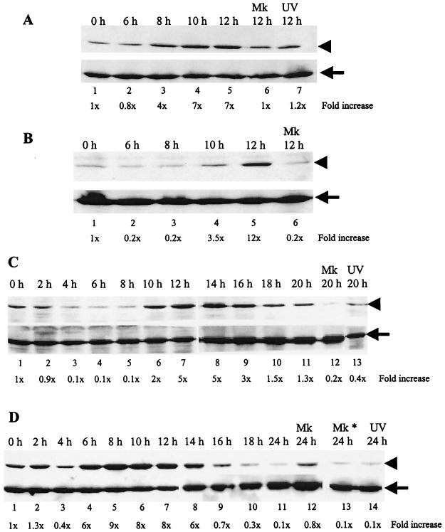
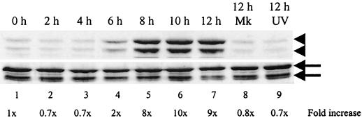
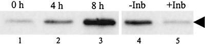
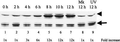
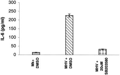
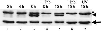
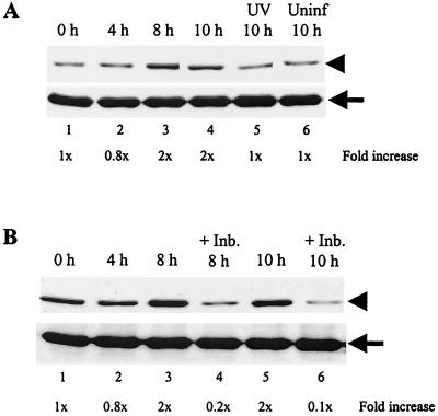
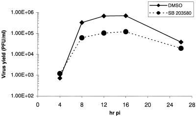
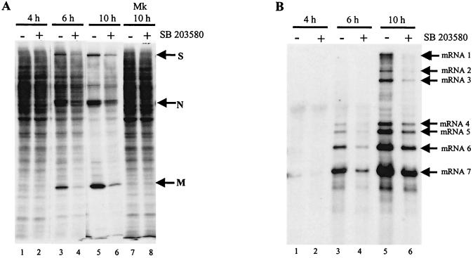
Analyses of mitogen-activated protein kinases (MAPKs) in a mouse hepatitis virus (MHV)-infected macrophage-derived J774.1 cell line showed activation of two MAPKs, p38 MAPK and c-Jun N-terminal kinase (JNK), but not of extracellular signal-regulated kinase (ERK). Activation of MAPKs was evident by 6 h postinfection. However, UV-irradiated MHV failed to activate MAPKs, which demonstrated that MHV replication was necessary for their activation. Several other MHV-permissive cell lines also showed activation of both p38 MAPK and JNK, which indicated that the MHV-induced stress-kinase activation was not restricted to any particular cell type. The upstream kinase responsible for activating MHV-induced p38 MAPK was the MAPK kinase 3. Experiments with a specific inhibitor of p38 MAPK, SB 203580, demonstrated that MHV-induced p38 MAPK activation resulted in the accumulation of interleukin-6 (IL-6) mRNAs and an increase in the production of IL-6, regardless of MHV-induced general host protein synthesis inhibition. Furthermore, MHV production was suppressed in SB 203580-treated cells, demonstrating that activated p38 MAPK played a role in MHV replication. The reduced MHV production in SB 203580-treated cells was, at least in part, due to a decrease in virus-specific protein synthesis and virus-specific mRNA accumulation. Interestingly, there was a transient increase in the amount of phosphorylation of the translation initiation factor 4E (eIF4E) in infected cells, and this eIF4E phosphorylation was p38 MAPK dependent; it is known that phosphorylated eIF4E enhances translation rates of cap-containing mRNAs. Furthermore, the upstream kinase responsible for eIF4E phosphorylation, MAPK-interacting kinase 1, was also phosphorylated and activated in response to MHV infection. Our data suggested that host cells, in response to MHV replication, activated p38 MAPK, which subsequently phosphorylated eIF4E to efficiently translate certain host proteins, including IL-6, during virus-induced severe host protein synthesis inhibition. MHV utilized this p38 MAPK-dependent increase in eIF4E phosphorylation to promote virus-specific protein synthesis and subsequent progeny virus production. Enhancement of virus-specific protein synthesis through virus-induced eIF4E activation has not been reported in any other viruses.


Figures

FIG. 1.
FIG. 1.
Phosphorylation of p38 MAPK in MHV-infected 17Cl-1 (A), L929 (B), DBT (C), and J774.1 (D) cells. Cells were infected with MHV at an MOI of 20 (A, C, and D) or 100 (B). Cell extracts were prepared at the indicated times p.i. Western blot analysis with a p38-phospho-specific antibody (upper panels in A to D) demonstrates p38 MAPK that is dually phosphorylated at residues Thr180/Tyr182, while all forms of p38 are detected by a p38 antibody that recognizes total p38 protein (bottom panels in A to D). Arrowheads, phosphorylated p38 MAPK; arrows, total p38 MAPK; h, h p.i.; Mk, mock-infected cells; Mk*, cells treated with UV-irradiated culture medium; UV, cells infected with the inoculum containing UV-irradiated MHV. The phospho-specific signal in each immunoblot was quantitated by densitometric scanning and normalized against each total protein. The increase in phosphorylation for each sample is denoted as the fold increase over the signal from the zero hour p.i. time point and is indicated below each lane.
FIG. 2.
FIG. 2.
JNK phosphorylation in MHV-infected cells. J774.1 cells were mock infected (Mk), MHV infected, or exposed to inoculum containing UV-irradiated MHV (UV). Cell lysates were harvested at the indicated times and analyzed by Western blot with a JNK phospho-specific antibody that detects JNK phosphorylated at residues Thr183 and Tyr185, shown in the upper panel (arrowheads), and a phosphorylation state-independent antibody that detects total JNK, shown in the lower panel (arrows). The upper arrow and upper arrowhead indicate JNK p54; the lower arrow and lower arrowhead indicate JNK p46.
FIG. 3.
FIG. 3.
Kinase activity of MHV-induced phosphorylated p38 in vitro. Cell extracts were prepared from MHV-infected J774.1 cells at the indicated times p.i. (lanes 1 to 3). For lanes 1 to 3, lysates were immunoprecipitated with p38-phospho-specific antibody and the p38 kinase activity of the immunecomplex was determined by using recombinant ATF-2 as the substrate in an in vitro kinase assay. Kinase reactions were analyzed by Western blot with an ATF-2-phospho-specifc antibody that detects ATF-2 phosphorylated at Thr71. For lanes 4 and 5, protein lysates that were extracted at 8 h p.i. were used for immunoprecipitation. “−Inb” and “+Inb” refer to immunoprecipitated complexes that were treated with DMSO and 20 μM SB 203580, respectively, prior to adding ATF-2 substrate. The arrowhead denotes phosphorylated ATF-2.
FIG. 4.
FIG. 4.
MKK3 phosphorylation in MHV-infected cells. J774.1 cells were mock infected (Mk), MHV infected, or exposed to inoculum containing UV-irradiated MHV (UV). Cell lysates were harvested at the indicated times and analyzed by Western blot with an MKK3/6 phospho-specific antibody that detects MKK3/6 phosphorylated at Ser189/207, shown in the upper panel (arrowhead), or with a phosphorylation-state-independent antibody that detects total MKK3 alone (not MKK6), shown in the lower panel (arrow).
FIG. 5.
FIG. 5.
MHV-induced p38 MAPK activation-dependent IL-6 secretion. Mock-infected J774.1 cells (Mk+DMSO) or MHV-infected J774.1 cells were incubated in the presence of DMSO (MHV+DMSO) or 20 μM SB 203580 (MHV+20 μM SB 203580). ELISA was used to determine the amount of IL-6 present in culture fluid at 24 h p.i. Error bars indicate the standard deviation from experimental points in triplicate determinations.
FIG. 6.
FIG. 6.
Mnk1 phosphorylation in MHV-infected cells. J774.1 cells were pretreated with DMSO or 20 μM SB 203580 (+Inb.) for 1 h prior to MHV infection. Then cells were infected with MHV or UV-irradiated MHV (UV) for 1 h. The virus inoculum was removed, and the cells were kept continuously in the presence of DMSO or SB 203580 (+Inb.). At the indicated times p.i., lysates were prepared and 30-μg protein lysates were analyzed by Western blot with a Mnk1 phospho-specific antibody, shown in the upper panel (arrowhead), that detects Mnk1 phosphorylated at Thr197/202 and with a phosphorylation-state-independent antibody, shown in the lower panel (arrow), that detects total Mnk1. The asterisk represents a nonspecific protein band.
FIG. 7.
FIG. 7.
Accumulation of phosphorylated eIF4E in MHV-infected cells. (A) J774.1 cells were left uninfected (Uninf), MHV infected, or exposed to inoculum containing UV-irradiated MHV (UV). At the times indicated, cell lysates were prepared and 30-μg protein lysates were analyzed by Western blot analysis with eIF4E phospho-specific antibody that detects eIF4E phosphorylated at Ser209, shown in the upper panel. The blots were subsequently stripped and reprobed with an eIF4E antibody that recognizes all forms of eIF4E (shown in the lower panel). The arrowhead and arrow denote phosphorylated eIF4E and total eIF4E, respectively. (B) J774.1 cells were pretreated with DMSO or 20 μM SB 203580 (+Inb.) for 1 h prior to MHV infection. Cells were then infected with MHV for 1 h. After the virus inoculum was removed, the cells were kept continuously in the presence of DMSO or SB 203580 (+Inb.). At the indicated times p.i., lysates were prepared and Western blot analysis was performed as described in panel A. The increase in the phospho-specific signal, normalized against the total protein signal and expressed as a fold increase over the zero hour p.i. signal, is indicated below each lane.
FIG. 8.
FIG. 8.
Effect of p38 MAPK activation on MHV production. J774.1 cells that were pretreated with DMSO or 20 μM SB 203580 for 1 h were infected with MHV for 1 h. After removal of the inoculum, the cells were continuously treated with DMSO (⧫) or 20 μM SB 203580 (•). The release of infectious MHV into the supernatant at the indicated times p.i. was determined by plaque assay.
FIG. 9.
FIG. 9.
Effect of p38 MAPK activity inhibition on translation (A) and virus-specific mRNA transcription (B). (A) J774.1 cells were left untreated (−) or were treated with 20 μM SB 203580 (+) for 1 h prior to infection. Cells were mock (Mk) or MHV infected. After 1 h of incubation, the inoculum was replaced with medium either lacking (−) or containing (+) 20 μM SB 203580. The culture medium was replaced with methionine-cysteine-free medium lacking (−) or containing (+) 20 μM SB 203580 1 h prior to each indicated time point. After 30 min of incubation, 75 μCi of Tran35S-label/ml was added to the culture medium. After 30 min of additional incubation, cell extracts were prepared and one-half of each sample was analyzed by SDS-12% PAGE. The positions of the MHV structural proteins S, N, and M are indicated by arrows. (B) Intracellular RNA was extracted from the remaining half of the lysates described above. After RNA was separated on a 1% agarose-formaldehyde gel, the RNAs were transferred to a nylon membrane. Northern blot analysis was performed with a random-primed DIG-labeled probe, specific for all MHV mRNAs. The positions of the MHV-specific mRNAs mRNA1 to mRNA7 are indicated with arrows.

Similar articles

Cited by

References

    1. An, S., C. J. Chen, X. Yu, J. L. Leibowitz, and S. Makino. 1999. Induction of apoptosis in murine coronavirus-infected cultured cells and demonstration of E protein as an apoptosis inducer. J. Virol. 73:7853-7859. - PMC - PubMed
    1. An, S., A. Maeda, and S. Makino. 1998. Coronavirus transcription early in infection. J. Virol. 72:8517-8524. - PMC - PubMed
    1. Banerjee, S., S. An, A. Zhou, R. H. Silverman, and S. Makino. 2000. RNase L-independent specific 28S rRNA cleavage in murine coronavirus-infected cells. J. Virol. 74:8793-8802. - PMC - PubMed
    1. Baric, R. S., S. A. Stohlman, and M. M. Lai. 1983. Characterization of replicative intermediate RNA of mouse hepatitis virus: presence of leader RNA sequences on nascent chains. J. Virol. 48:633-640. - PMC - PubMed
    1. Chang, L., and M. Karin. 2001. Mammalian MAP kinase signalling cascades. Nature 410:37-40. - PubMed

Publication types

MeSH terms

Substances