. 2002 Jun;76(12):5966-73.
doi: 10.1128/jvi.76.12.5966-5973.2002.

Effects of antiretroviral drugs on human immunodeficiency virus type 1-induced CD4(+) T-cell death

Affiliations

Effects of antiretroviral drugs on human immunodeficiency virus type 1-induced CD4(+) T-cell death

Jérôme Estaquier et al. J Virol. 2002 Jun.

Abstract

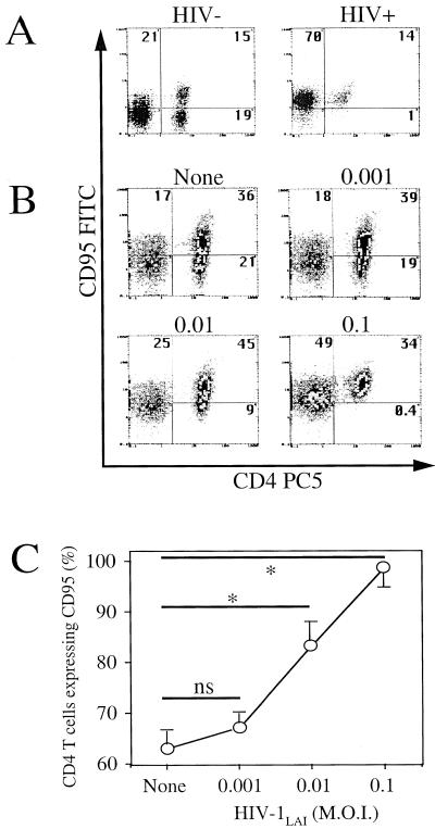
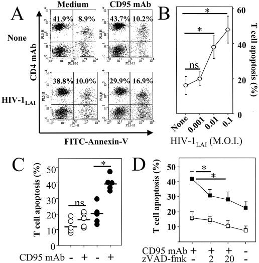
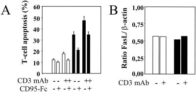
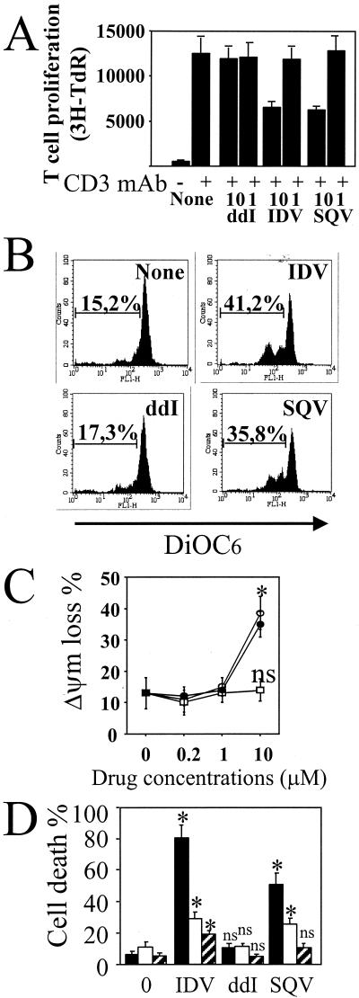
Apoptosis of peripheral blood T cells plays an important role in the pathogenesis of human immunodeficiency virus (HIV) infection. In this study, we found that HIV type 1 (HIV-1) primes CD4(+) T cells from healthy donors for apoptosis, which occurs after CD95 ligation or CD3-T-cell receptor (TCR) stimulation. CD95-mediated death did not depend on CD4 T-cell infection, since it occurred in the presence of the reverse transcriptase inhibitor didanosine (ddI). In contrast, apoptosis induced by productive infection (CD3-TCR stimulation) is prevented by both CD95 decoy receptor and ddI. Our data suggest that HIV-1 triggers at least two distinct death pathways: a CD95-dependent pathway that does not require viral replication and a viral replication-mediated cell death independent of the CD95 pathway. Further experiments indicated that saquinavir, a protease inhibitor, at a 0.2 microM concentration, decreased HIV-mediated CD95 expression and thus cell death, which is independent of its role in inhibiting viral replication. However, treatment of peripheral blood mononuclear cells from healthy donors with a higher concentration (10 microM) of an HIV protease inhibitor, saquinavir or indinavir, induced both a loss in mitochondrial membrane potential (DeltaPsim) and cell death. Thus, protease inhibitors have the potential for both beneficial and detrimental effects on CD4(+) T cells independent of their antiretroviral effects.


Figures

FIG. 1.
FIG. 1.
HIV-1LAI-induced CD95 expression. (A) CD95 expression in CD4+ T cells of an uninfected individual (HIV−) and an HIV-infected (HIV+) individual assessed by two-color flow cytometry using CD4- and CD95-labeled antibodies. Numbers indicate percentages in each quadrant. (B) PBMC from a healthy donor were incubated for 2 h in the absence (None) or presence of HIV-1LAI at different MOIs (0.001, 0.01, or 0.1) and then washed and further incubated for 4 days in medium alone. CD95 expression was assessed in CD4+ T cells by two-color flow cytometry. Numbers indicate percentages in each quadrant. (C) The proportion of CD4+ T cells expressing CD95 was calculated as follows: [CD4+ CD95+/(CD4+ CD95+ + CD4+ CD95)] × 100. Results are the means ± standard deviations of four independent experiments. Statistical significance was assessed by Student's t test (∗, P < 0.05; ns, no significant statistical difference).
FIG. 2.
FIG. 2.
HIV-1LAI-mediated priming of CD4+ T cells for CD95-induced death. (A) PBMC were incubated for 2 h in the absence (None) or presence of HIV-1LAI (MOI of 0.01), and cells were washed and incubated for 4 days in medium alone (Medium) or in the presence of an agonistic CD95 MAb (1 μg/ml) (CD95 MAb). Numbers indicate percentages in each quadrant. Dying CD4+ T cells were assessed by two-color flow cytometry, with PC5-labeled CD4 MAb and FITC-labeled annexin V. (B) The percentage of dying CD4+ T cells was calculated as follows: [CD4+ annexin+/(CD4+ annexin+ + CD4+ annexin)] × 100. Results are the means ± standard deviations of three independent experiments. Statistical significance was assessed by Student's t test (∗, P < 0.05; ns, no significant statistical difference). (C) Purified CD4+ T cells were incubated for 2 h in the absence (○) or presence (•) of HIV-1LAI (MOI of 0.01) and then washed and incubated for 4 days in medium alone (−) or in the presence (+) of an agonistic CD95 IgM MAb (1 μg/ml). Percentages of dying CD4+ T cells were assessed by two-color flow cytometry, with CD4 MAb and annexin V, and were calculated as follows: [CD4+ annexin+/(CD4+ annexin+ + CD4+ annexin)] × 100. Each symbol represents one individual donor, and bars represent mean values in each group. Statistical significance was assessed by Student's t test (∗, P < 0.05; ns, no significant statistical difference). (D) Caspase inhibitor decreased CD95-mediated T-cell death. CD4+ T cells were incubated for 2 h in the absence (□) or presence (▪) of HIV-1LAI (MOI of 0.01) and then washed and further incubated in medium alone for 4 days. CD4+ T cells were then further incubated for 18 h in the absence (−) or presence (+) of 1 μg of agonistic anti-CD95 (CD95 MAb) per ml and in the absence (−) or presence (+) of a 2 μM (“2”) or a 20 μM (“20”) concentration of the broad caspase inhibitor zVAD-fmk. Percentages of dying CD4+ T cells were assessed by flow cytometry with annexin V. The percentage of dying CD4+ T cells was calculated as follows: [CD4+ annexin+/(CD4+ annexin+ + CD4+ annexin)] × 100. Results are the means ± standard deviations of three independent experiments. Statistical significance was assessed by Student's t test (∗, P < 0.05).
FIG. 3.
FIG. 3.
HIV-1LAI-mediated priming of CD4+ T cells for CD3-induced death. (A) HIV-1-mediated priming of CD4+ T cells for CD3-induced T-cell death. PBMC were incubated for 2 h in the absence (open bars) or presence (solid bars) of HIV-1LAI (at an MOI of 0.01) and then washed and further incubated in medium alone for 4 days. CD4+ T cells were then purified, by negative selection, and then cultured for 18 h in the absence (−) or presence (+) of CD3 MAb (1 μg/ml) and in the absence (−) or presence (+) of a soluble CD95 decoy receptor (human CD95-Fc Ig fusion protein) (10 μg/ml). Percentages of dying CD4+ T cells were assessed by flow cytometry with annexin V and were calculated as follows: [CD4+ annexin+/(CD4+ annexin+ + CD4+ annexin)] × 100. The results for one typical experiment out of three performed in duplicate (means ± standard deviations) are shown. (B) Expression of CD95L mRNA was assessed by an RNase protection assay in purified CD4+ T cells (107 cells) isolated from PBMC incubated for 4 days earlier in the absence (open bars) or presence (solid bars) of HIV-1LAI. CD4+ T cells were then further incubated for 6 h in the absence (−) or presence (+) of CD3 MAb (1 μg/ml).
FIG. 4.
FIG. 4.
Effects of HIV drugs on PBMC from healthy donors. (A) PBMC were incubated in the absence (None) or presence of ddI, SQV, and IDV, at different concentrations (10 and 1 μM). After 1 h of in vitro treatment, PBMC were stimulated with CD3 MAb (1 μg/ml). T-cell proliferation was assessed after 3 days. Data represent one typical experiment out of three performed in triplicate (means ± standard deviations). TdR, thymidine. (B) Loss in mitochondrial membrane potential (ΔΨm) in dying cells. ΔΨm was assessed by flow cytometry with DiOC6 at day 3 from unstimulated PBMC. Numbers indicate percentages of ΔΨm loss. PBMC were incubated in the absence (None) or presence of ddI (10 μM), SQV (10 μM), and IDV (10 μM). (C) PBMC were incubated in the absence (0 μM) or presence of ddI (□), SQV (•), and IDV (○), at different concentrations (10, 1, and 0.2 μM). Results are the means of three independent experiments (means ± standard deviations). Statistical significance was calculated by comparing the percentage of ΔΨm loss in treated cells with the percentage in the untreated cells by Student's t test (∗, P < 0.05; ns, no significant statistical difference). (D) PBMC were incubated either in the absence (0) or in the presence of ddI, SQV, and IDV at 10 μM. Specific monocyte (solid bars), CD4 (open bars), and CD8 (hatched bars) T-cell death was assessed after 3 days by two-color flow cytometry with annexin V. Results are the means of three independent experiments (means ± standard deviations). Statistical significance was calculated by comparing the percentage of annexin V-positive cells in treated culture of a particular phenotype with the percentage in the untreated culture by Student's t test (∗, P < 0.05; ns, no significant statistical difference).
FIG. 5.
FIG. 5.
HIV drugs decreased CD95 and CD3-mediated cell death. (A) Schedule of experiment. (B) PBMC were incubated for 2 h in the absence (open bars) or presence (solid bars) of HIV-1LAI (MOI of 0.01), and cells were washed and incubated in the absence (None) or presence of ddI (5 μM) and SQV (0.2 μM). CD4+ T cells were purified and then cultured for 18 h in the absence (Medium) or presence of CD95 MAb (1 μg/ml). Results are the means of three independent experiments (means ± standard deviations). Statistical significance was assessed by Student's t test (∗, P < 0.05; ns, no significance). (C) CD95 expression of CD4+ T cells from PBMC of healthy donors incubated for 2 h in the absence (open bars) or presence (solid bars) of HIV-1LAI (MOI of 0.01), for 4 days in medium alone (None) or in the presence of drugs (ddI and SQV). CD95 expression was assessed in CD4+ T cells by two-color flow cytometry. The proportion of CD4+ T cells expressing CD95 was calculated as follows: [CD4+ CD95+/(CD4+ CD95+ + CD4+ CD95)] × 100. Results are the means of three independent experiments (means ± standard deviations). Statistical significance was assessed by Student's t test (∗, P < 0.05; ns, no significance). (D) CD4+ T cells were isolated as described for panel B and further cultured for 18 h in the presence of CD95 MAb (1 μg/ml) or CD3 MAb (1 μg/ml) in the absence (CD3 and CD95) or presence of IL-2 (CD3 IL-2 and CD95 IL-2) (20 ng/ml). Cell death was assessed by flow cytometry with annexin V. The percentage of dying CD4+ T cells was calculated as follows: [CD4+ annexin+/(CD4+ annexin+ + CD4+ annexin)] × 100. Results are the means of two independent experiments (means ± standard deviations).
FIG. 6.
FIG. 6.
T-cell proliferation is restored in the presence of ddI and CD95 decoy receptor. CD4+ T cells were incubated with HIV-1LAI and purified as described for Fig. 5B. Cells were incubated in the absence (open bars) or presence (solid bars) of 5 μM ddI added at day 0 and day 2. CD4+ T cells were further stimulated in the absence (Med) or presence of CD3 MAb (1 μg/ml) or CD3 with CD28 MAbs (CD3 CD28) (1 μg/ml each). At day 3, T-cell proliferation (A), T-cell counts (B), and p24 antigen production (C) were assessed. (A) T-cell proliferation was determined by [3H]thymidine (TdR) incorporation. Results are the means of three independent experiments (means ± standard deviations). (B) T-cell counts were assessed by light microscopy. Results are the means of three independent experiments (means ± standard deviations). (C) Enzyme-linked immunosorbent assay specific for p24 antigen was used to assess viral production. Results are the means of three independent experiments (means ± standard deviations). Statistical significance was assessed by Student's t test (∗, P < 0.05; ns, no significant statistical difference). (D) PBMC were incubated for 2 h in the absence (open bars) or presence (filled bars) of HIV-1LAI (at an MOI of 0.01) and then washed and further incubated in medium alone (None) or in the presence of ddI for 4 days. CD4+ T cells then were purified, by negative selection, and cultured for 18 h inthe presence of either CD3 MAb (1 μg/ml) or CD3 and CD28 MAbs (1 μg/ml each). CD4+ T cells treated with ddI were also incubated in the presence of a soluble CD95 decoy receptor (ddI/CD95-Fc) (10 μg/ml). T-cell counts were assessed by light microscopy at day 4. Results are the means of two independent experiments (means ± standard deviations).

References

    1. Ameisen, J. C., J. Estaquier, and T. Idziorek. 1994. From AIDS to parasite infection: pathogen-mediated subversion of programmed cell death as a mechanism for immune dysregulation. Immunol. Rev. 142:9-51. - PubMed
    1. Amendola, A., F. Poccia, F. Martini, C. Gioia, V. Galati, M. Pierdominici, M. Marziali, F. Pandolfi, V. Colizzi, M. Piacentini, E. Girardi, G. D'Offizi, et al. 2000. Decreased CD95 expression on naive T cells from HIV-infected persons undergoing highly active anti-retroviral therapy (HAART) and the influence of IL-2 low dose administration. Clin. Exp. Immunol. 120:324-332. - PMC - PubMed
    1. Andre, P., M. Groettrup, P. Klenerman, R. de Giuli, B. L. Booth, Jr., V. Cerundolo, M. Bonneville, F. Jotereau, R. M. Zinkernagel, and V. Lotteau. 1998. An inhibitor of HIV-1 protease modulates proteasome activity, antigen presentation, and T cell responses. Proc. Natl. Acad. Sci. USA 95:13120-13124. - PMC - PubMed
    1. Aries, S. P., K. Weyrich, B. Schaaf, F. Hansen, R. H. Dennin, and K. Dalhoff. 1998. Early T-cell apoptosis and Fas expression during antiretroviral therapy in individuals infected with human immunodeficiency virus-1. Scand. J. Immunol. 48:86-91. - PubMed
    1. Badley, A. D., D. H. Dockrell, A. Algeciras, S. Ziesmer, A. Landay, M. M. Lederman, E. Connick, H. Kessler, D. Kuritzkes, D. H. Lynch, P. Roche, H. Yagita, and C. V. Paya. 1998. In vivo analysis of Fas/FasL interactions in HIV-infected patients. J. Clin. Investig. 102:79-87. - PMC - PubMed

Publication types

MeSH terms