. 2002 Jun;76(12):5974-84.
doi: 10.1128/jvi.76.12.5974-5984.2002.

Expression of hepatitis C virus proteins induces distinct membrane alterations including a candidate viral replication complex

Affiliations

Expression of hepatitis C virus proteins induces distinct membrane alterations including a candidate viral replication complex

Denise Egger et al. J Virol. 2002 Jun.

Abstract

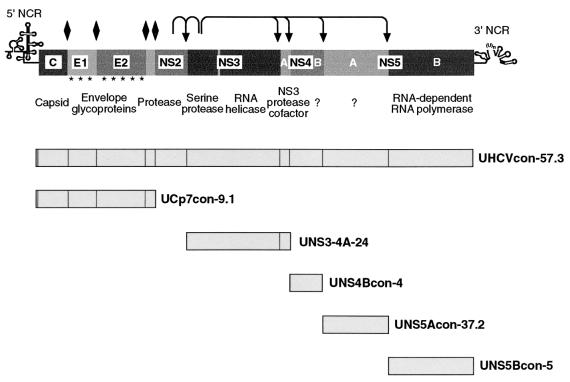
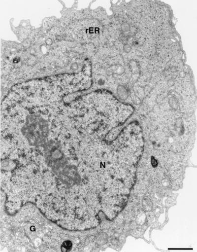
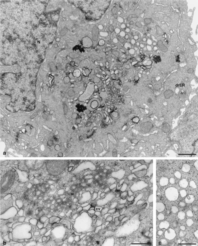
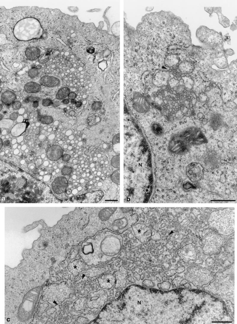
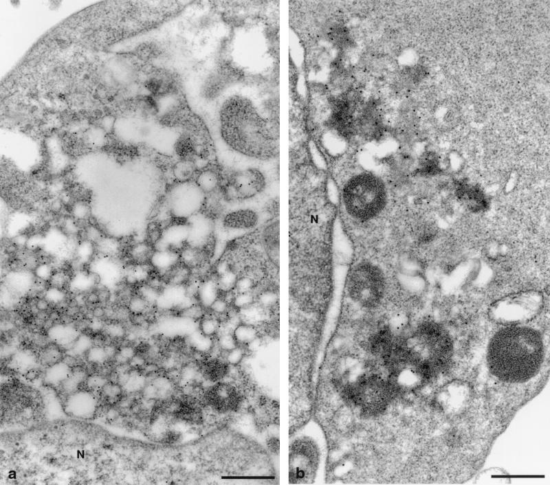
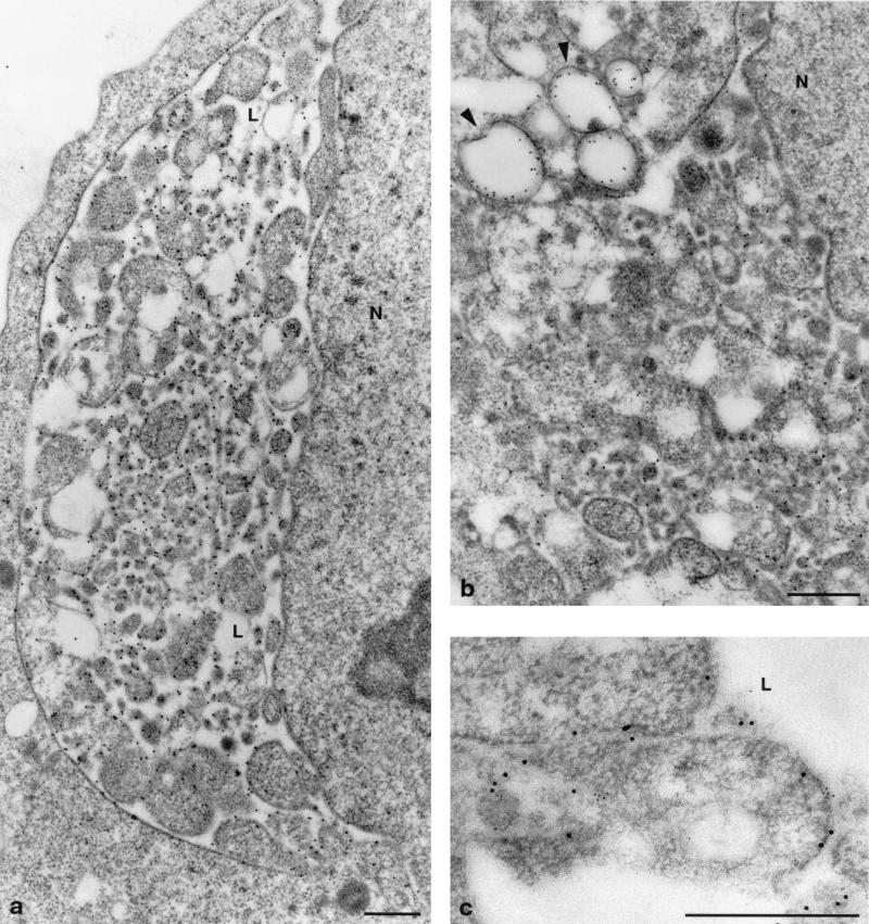
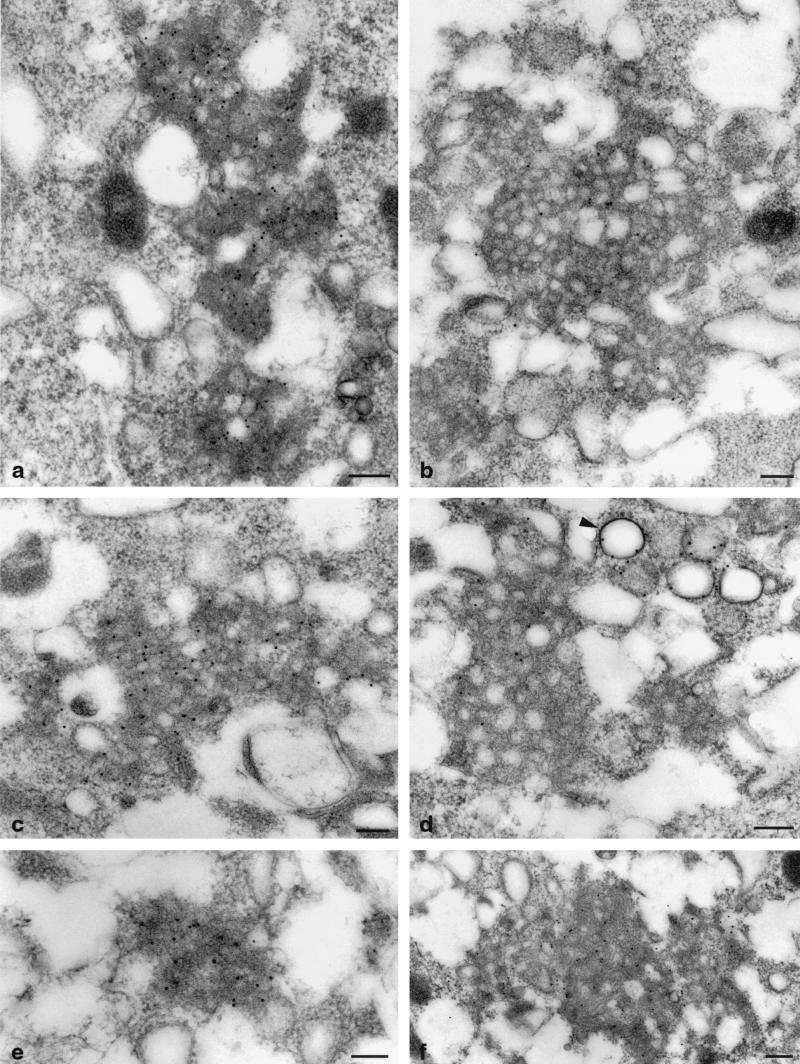
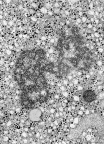
Plus-strand RNA viruses characteristically replicate their genome in association with altered cellular membranes. In the present study, the capacity of hepatitis C virus (HCV) proteins to elicit intracellular membrane alterations was investigated by expressing, in tetracycline-regulated cell lines, a comprehensive panel of HCV proteins individually as well as in the context of the entire HCV polyprotein. As visualized by electron microscopy (EM), expression of the combined structural proteins core-E1-E2-p7, the NS3-4A complex, and protein NS4B induced distinct membrane alterations. By immunogold EM (IEM), the membrane-altering proteins were always found to localize to the respective altered membranes. NS4B, a protein of hitherto unknown function, induced a tight structure, designated membranous web, consisting of vesicles in a membranous matrix. Expression of the entire HCV polyprotein gave rise to membrane budding into rough endoplasmic reticulum vacuoles, to the membranous web, and to tightly associated vesicles often surrounding the membranous web. By IEM, all HCV proteins were found to be associated with the NS4B-induced membranous web, forming a membrane-associated multiprotein complex. A similar web-like structure in livers of HCV-infected chimpanzees was previously described (Pfeifer et al., Virchows Arch. B., 33:233-243, 1980). In view of this finding and the observation that all HCV proteins accumulate on the membranous web, we propose that the membranous web forms the viral replication complex in HCV-infected cells.


Figures

FIG. 1.
FIG. 1.
Tetracycline-regulated cell lines. The genetic organization and polyprotein processing of HCV are depicted at the top. Asterisks in the E1 and E2 regions indicate glycosylation of the envelope proteins. Diamonds indicate cleavages of the HCV polyprotein precursor by the ER signal peptidase, and arrows indicate cleavages by HCV NS2-3 and NS3 proteases. The well-characterized and representative clones UHCVcon-57.3, UCp7con-9.1, UNS3-4A-24, UNS4Bcon-4, UNS5Acon-37.2, and UNS5Bcon-5 were used in this study.
FIG. 2.
FIG. 2.
UHCVcon-57.3 cell from a culture grown in the presence of tetracycline. The expression of the HCV polyprotein is repressed, and the cell shows no pathological findings. G, Golgi; rER, rough endoplasmic reticulum; N, nucleus. Bar, 1 μm.
FIG. 3.
FIG. 3.
UHCVcon-57.3 cells expressing the entire HCV polyprotein 48 h after tetracycline withdrawal. (a) Low-power overview showing the alterations induced. w, membranous web; li, lipid droplets; cv, contiguous vesicles; drER, dilated rER forming vacuoles; N, nucleus. Bar, 1 μm. (b) Higher magnification of the membranous web (w), surrounded by clusters of contiguous vesicles (cv). Bar, 0.5 μm. (c) Vacuoles of dilated rER, carrying ribosomes on their surfaces. Bar, 0.5 μm.
FIG. 4.
FIG. 4.
Cells expressing individual HCV proteins. (a) UNS3-4A-24 cells, expressing the proteins NS3 and NS4A, show smooth-surfaced vesicles of varying size. The vesicles are distributed in clusters in the cytoplasm. (b) Protein NS4B expressed by UNS4Bcon-4 cells induces a membranous web. Note the association of the web with remnants of the rER (arrowhead). (c) UCp7con-9.1 cells express the structural proteins core, E1, E2, and p7. Highly dilated rER vacuoles contain numerous invaginations of the rER, budding into the lumen of the large vacuole. Arrowheads, small buds; asterisks, large buds; N, nucleus. Bars, 0.5 μm.
FIG. 5.
FIG. 5.
Immunogold detection of HCV nonstructural proteins expressed individually. The viral proteins are associated with the membrane alterations which they induce. (a) UNS3-4A-24 cells labeled with anti-NS3 Ab. NS3 is found on smooth small vesicles. (b) UNS4Bcon-4 cells labeled with anti-NS4B Ab. NS4B is found exclusively on the membranous web. N, nucleus. Bars, 0.5 μm.
FIG. 6.
FIG. 6.
Immunogold detection of structural proteins expressed in UCp7con-9.1 cells. (a) E1 protein is found on the budding membrane invaginations in the lumen of the rER. (b) Core protein is also found on membranes in the lumen of the rER and, in addition, on lipid droplets (arrowheads). (c) Double immunogold staining shows proteins E1 (larger gold grains) and core (smaller gold grains) on the same membrane structures, albeit not closely associated with each other. L, lumen of rER; N, nucleus. Bars, 0.5 μm.
FIG. 7.
FIG. 7.
Immunogold detection of structural and nonstructural proteins in UHCVcon-57.3 cells expressing the entire HCV polyprotein. The cells were labeled with Abs against proteins NS3 (a), NS4B (b), NS5A (c), core (d), NS4A (e), and E1 (f). All proteins tested are found on the membranous web (a to f); the core protein is additionally found on lipid droplets (arrowhead) (d). Bars, 0.2 μm.
FIG. 8.
FIG. 8.
The liver biopsy of an HCV-infected chimpanzee shows a web-like inclusion whose size and structure are comparable to those found in UHCVcon-57.3 cells. G, glycogen granules. Bar, 0.5 μm.

Similar articles

Cited by

References

    1. Aldabe, R., and L. Carrasco. 1995. Induction of membrane proliferation by poliovirus proteins 2C and 2BC. Biochem. Biophys. Res. Commun. 206:64-76. - PubMed
    1. Barba, G., F. Harper, T. Harada, M. Kohara, S. Goulinet, Z. Matsuura, G. Eder, Z. Schaff, M. J. Chapman, T. Miyamura, and C. Bréchot. 1997. Hepatitis C virus core protein shows a cytoplasmic localization and associates to cellular lipid storage droplets. Proc. Natl. Acad. Sci. USA 94:1200-1205. - PMC - PubMed
    1. Bartenschlager, R., and V. Lohmann. 2000. Replication of hepatitis C virus. J. Gen. Virol. 81:1631-1648. - PubMed
    1. Behrens, S. E., L. Tomei, and R. Defrancesco. 1996. Identification and properties of the RNA-dependent RNA polymerase of hepatitis C virus. EMBO J. 15:12-22. - PMC - PubMed
    1. Bienz, K., and D. Egger. 1995. Immunocytochemistry and in situ hybridization in the electron microscope: combined application in the study of virus-infected cells. Histochem. Cell Biol. 103:325-338. - PubMed

Publication types