. 2002 Jun;76(12):6235-43.
doi: 10.1128/jvi.76.12.6235-6243.2002.

Coalescence of the sites of cowpea mosaic virus RNA replication into a cytopathic structure

Affiliations

Coalescence of the sites of cowpea mosaic virus RNA replication into a cytopathic structure

Jan E Carette et al. J Virol. 2002 Jun.

Abstract

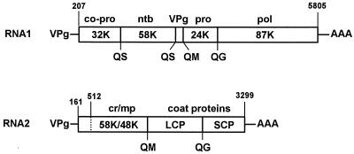
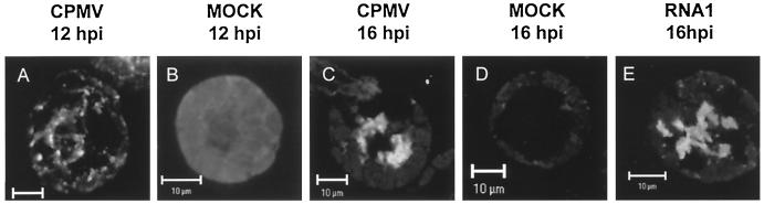
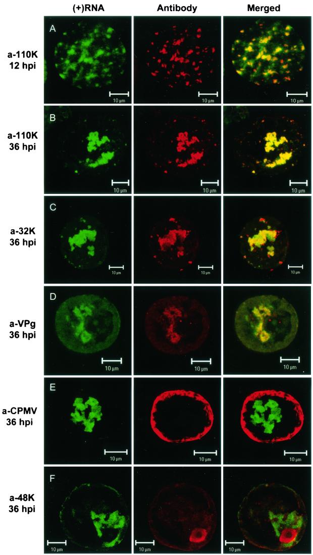
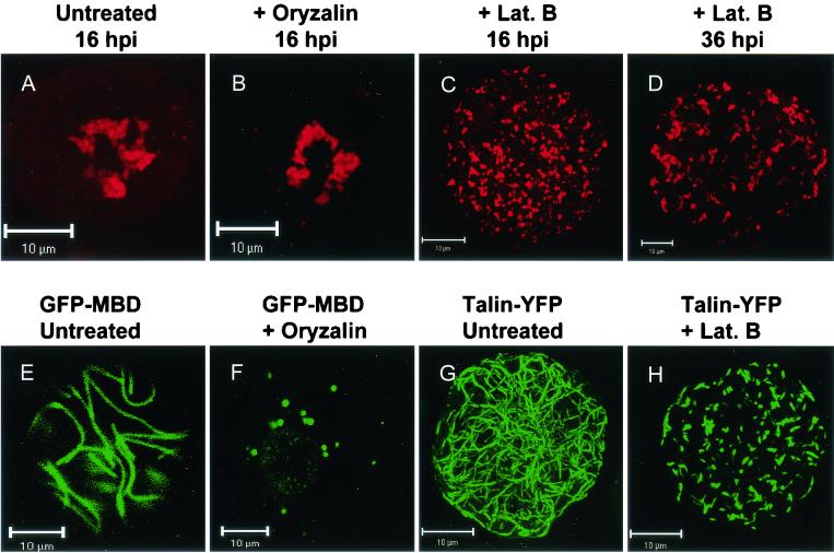
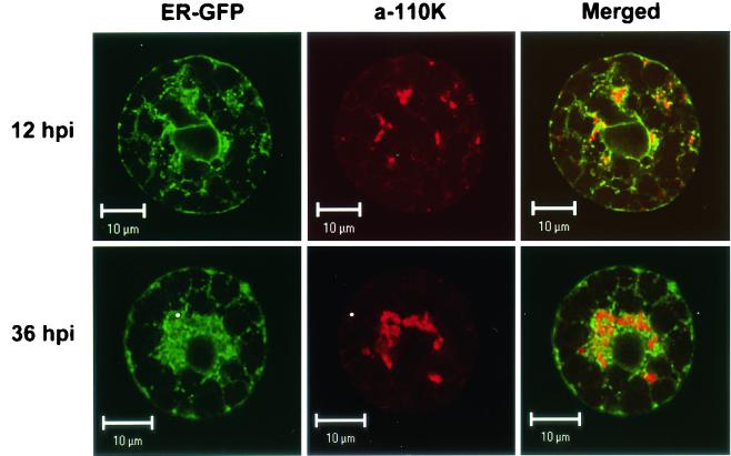
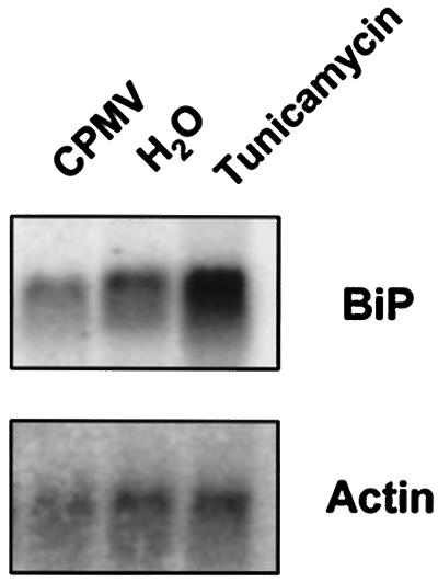
Cowpea mosaic virus (CPMV) replication induces an extensive proliferation of endoplasmic reticulum (ER) membranes, leading to the formation of small membranous vesicles where viral RNA replication takes place. Using fluorescent in situ hybridization, we found that early in the infection of cowpea protoplasts, CPMV plus-strand RNA accumulates at numerous distinct subcellular sites distributed randomly throughout the cytoplasm which rapidly coalesce into a large body located in the center of the cell, often near the nucleus. The combined use of immunostaining and a green fluorescent protein ER marker revealed that during the course of an infection, CPMV RNA colocalizes with the 110-kDa viral polymerase and other replication proteins and is always found in close association with proliferated ER membranes, indicating that these sites correspond to the membranous site of viral replication. Experiments with the cytoskeleton inhibitors oryzalin and latrunculin B point to a role of actin and not tubulin in establishing the large central structure. The induction of ER membrane proliferations in CPMV-infected protoplasts did not coincide with increased levels of BiP mRNA, indicating that the unfolded-protein response is not involved in this process.


Figures

FIG. 1.
FIG. 1.
Genetic organization of the CPMV genome. Open reading frames in the RNA molecules are indicated by open bars. The nucleotide positions of the start and stop codons are shown above the open reading frame, and the cleavage sites in the polyproteins are shown below. The 110K (24K plus 87K) intermediate processing product is the full-length polymerase (13). Abbreviations: co-pro, cofactor for proteinase; ntb, nucleotide-binding protein; pro, proteinase; pol, core polymerase; cr, cofactor for RNA2 replication; mp, movement protein; LCP, large coat protein; SCP, small coat protein.
FIG. 2.
FIG. 2.
Intracellular distribution of plus-strand viral RNA in protoplasts infected with CPMV RNA. Protoplasts were collected at the indicated time points after infection with CPMV RNA1 and RNA2 (CPMV), CPMV RNA1 (RNA1), or water (mock) and hybridized with a fluorescein-RNA probe that recognized plus-strand CPMV RNA1. Fluorescent signals were visualized by confocal microscopy measuring optical sections with a focal depth of 1 μm. (A) At 12 hpi, viral RNA was localized in multiple small bodies dispersed over the cytoplasm. (C and E) At 16 hpi, viral RNA was observed in one or several large, amorphous bodies of fluorescence located in the center of protoplasts infected with CPMV (C) or with CPMV RNA1 alone (E). (B and D) Background fluorescence was evaluated in mock-infected protoplasts observed with identical settings at 12 hpi (B) and 16 hpi (D). Bars, 10 μm.
FIG.3.
FIG.3.
Localization of plus-strand viral RNA with respect to different viral proteins in CPMV-infected protoplasts. Protoplasts were collected at the indicated time points after infection with CPMV RNA, hybridized with a fluorescein-RNA probe that recognized plus-strand CPMV RNA1 (green signal), and immunostained with the indicated antisera (red signal). (A and B) At 12 and 36 hpi, the 110K polymerase was localized almost exclusively in sites where viral RNA accumulated. Colocalization of the two signals is shown in the merged images as yellow. (C) The 32K cofactor for the proteinase also colocalized substantially with viral RNA. (D) The majority of the proteins recognized by anti-VPg (a-VPg) serum colocalized with viral RNA. (E) Viral particles localized in the periphery of the cell and did not colocalize with the sites of viral RNA accumulation. (F) The 58K protein recognized by the anti-48K/58K serum localized mainly in the nucleus and did not colocalize with viral RNA. Bars, 10 μm.
FIG. 4.
FIG. 4.
Disruption of the actin but not the microtubular network induced changes in the localization of the viral 110K polymerase in CPMV-infected protoplasts. Immediately after infection, protoplasts were treated with oryzalin (10 μM) (B) or latrunculin B (20 μM) (C and D) or left untreated (A) and harvested at the indicated time points, followed by immunostaining with anti-110K serum. In untreated (A) and oryzalin-treated (B) protoplasts, the polymerase was present in a central large amorphous structure at 16 hpi. In cells treated with latrunculin B, the polymerase was present in numerous small spots scattered throughout the cytoplasm at both 16 hpi (C) and 36 hpi (D). Protoplasts transiently expressing GFP-MBD to visualize the microtubules (E and F) or Talin-YFP to visualize the actin cytoskeleton (G and H) show that oryzalin (F) and latrunculin B (H) were active at the concentration used at 16 h posttransfection. Lat. B, latrunculin B. Bars, 10 μm.
FIG. 5.
FIG. 5.
Dual localization of ER-targeted GFP (ER-GFP) and the viral 110K polymerase (a-110K) in CPMV-infected protoplasts. Cells were fixed at 12 hpi (top row) and 36 hpi (bottom row) and processed for immunofluorescence by using antibodies raised against the 110K polymerase. ER-GFP retained its fluorescence throughout the procedure. Proliferated ER membranes surrounded and traversed the sites of 110K accumulation both early (12 hpi) and late (36 hpi) in infection. Bars, 10 μm.
FIG. 6.
FIG. 6.
Effect of CPMV infection on BiP mRNA levels in cowpea protoplasts. Cowpea protoplasts were infected with CPMV (CPMV) or uninfected (H2O and Tunicamycin). Prior to the harvesting of the protoplasts (36 hpi), the sample designated Tunicamycin was treated with tunicamycin (20 μg/ml) for 2.5 h. Total RNA was extracted, separated on an agarose gel, and blotted on a nylon membrane. The blots were probed for BiP, and an actin probe (Actin) was used as a control for loading differences. CPMV infection (CPMV) did not lead to an increase in BiP mRNA levels compared to those of the uninfected protoplasts (H2O). Treatment with tunicamycin (Tunicamycin) resulted in upregulation of BiP expression.

References

    1. Argos, P., G. Kamer, M. J. Nicklin, and E. Wimmer. 1984. Similarity in gene organization and homology between proteins of animal picornaviruses and a plant comovirus suggest common ancestry of these virus families. Nucleic Acids Res. 12:7251-7267. - PMC - PubMed
    1. Assink, A. M., H. Swaans, and A. Van Kammen. 1973. The localization of virus-specific double-stranded RNA of cowpea mosaic virus in subcellular fractions of infected Vigna leaves. Virology 53:384-391. - PubMed
    1. Baluska, F., J. Jasik, H. G. Edelmann, T. Salajova, and D. Volkmann. 2001. Latrunculin B-induced plant dwarfism: plant cell elongation is F-actin-dependent. Dev. Biol. 231:113-124. - PubMed
    1. Baskin, T. I., J. E. Wilson, A. Cork, and R. E. Williamson. 1994. Morphology and microtubule organization in Arabidopsis roots exposed to oryzalin or taxol. Plant Cell Physiol. 35:935-942. - PubMed
    1. Beachy, R. N., and M. Heinlein. 2000. Role of P30 in replication and spread of TMV. Traffic 1:540-544. - PubMed

Publication types

LinkOut - more resources