. 2002 Jun;76(12):6268-76.
doi: 10.1128/jvi.76.12.6268-6286.2002.

1-beta-D-arabinofuranosylcytosine inhibits borna disease virus replication and spread

Affiliations
Free PMC article

1-beta-D-arabinofuranosylcytosine inhibits borna disease virus replication and spread

Jeffrey J Bajramovic et al. J Virol. 2002 Jun.
Free PMC article

Abstract

Borna disease virus (BDV) is a nonsegmented, negative-strand RNA virus that causes neurological diseases in a variety of warm-blooded animal species. There is general consensus that BDV can also infect humans, being a possible zoonosis. Although the clinical consequences of human BDV infection are still controversial, experimental BDV infection is a well-described model for human neuropsychiatric diseases. To date, there is no effective treatment against BDV. In this paper, we demonstrate that the nucleoside analog 1-beta-D-arabinofuranosylcytosine (Ara-C), a known inhibitor of DNA polymerases, inhibits BDV replication. Ara-C treatment inhibited BDV RNA and protein synthesis and prevented BDV cell-to-cell spread in vitro. Replication of other negative-strand RNA viruses such as influenza virus or measles virus was not inhibited by Ara-C, underscoring the particularity of the replication machinery of BDV. Strikingly, Ara-C treatment induced nuclear retention of viral ribonucleoparticles. These findings could not be attributed to known effects of Ara-C on the host cell, suggesting that Ara-C directly inhibits the BDV polymerase. Finally, we show that Ara-C inhibits BDV replication in vivo in the brain of infected rats, preventing persistent infection of the central nervous system as well as the development of clinical disease. These findings open the way to the development of effective antiviral therapy against BDV.


Figures
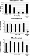
FIG. 1.
FIG. 1.
The effects of Ara-C and ribavirin on the titers of cell-free BDV, influenza virus, and measles virus. (Top) BDV cell-free viral titers, in FFU per milliliter of isolate. (Middle) Influenza virus strain Sydney (░⃞) and strain Puerto Rico (▪) viral titers in PFU are given as the percentages of untreated controls. (Bottom) Measles virus strain Edmonston B viral titers (50% tissue culture infective dose; Kärber method) are given as the percentages of untreated controls.
FIG. 2.
FIG. 2.
Ara-C inhibits BDV cell-to-cell spread. Confluent Vero cells were labeled with CFDA and subsequently cocultivated for 5 days at a ratio of 1:1 with unlabeled, BDV-infected Vero cells (Vero-BV). Cocultivation took place under daily treatment with different doses of Ara-C or with 20 μM ribavirin, after which cells were analyzed by flow cytometry. The percentages shown indicate the double-positive population within the population of viable cells, which indicates the amount of viral spread. The negative control consisted of a 1:1 mix of CFDA-labeled Vero cells with Vero-BV cells, fixed directly after mixing.
FIG. 3.
FIG. 3.
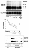
BDV RNA and protein expression levels are inhibited by Ara-C. Total cellular RNA and proteins from BDV-infected Vero cells were extracted at day 5 of treatment and analyzed. (Top) Northern blot analysis. The expected sizes of the different BDV mRNAs and genomic RNA are indicated. (Middle) Quantitation of BDV mRNA and genomic BDV RNA levels was done by PhosphorImager analysis and was thereafter standardized to GAPDH levels. Results are expressed as the percentages of untreated control levels, which were set to 100%. ○, genomic RNA; □, mRNA of 3.5 kb; ▴, mRNA of 1.9 kb; and ×, mRNA of 1.2 kb. (Bottom) Western blot analysis. Equal amounts of protein were resolved by SDS-PAGE, transferred to a Hybond membrane, and reacted with anti-N and anti-P antibodies (as indicated).
FIG. 4.
FIG. 4.
The subcellular distribution of BDV viral proteins is dramatically changed by Ara-C treatment. (Top) Vero-BV cells at day 5 of treatment were stained with anti-N antibody followed by FITC-conjugated anti-rabbit antibody. Original magnification, ×200. Similar results were obtained with C6 and U373 cells as well as with the 38/17C1 monoclonal antibody against N or a polyclonal antibody against BDV-P (data not shown). (Bottom) Cell fractionation followed by Western blot analysis. Vero-BV cells at day 5 of treatment were lysed and lysates were separated into nuclear and cytoplasmic fractions. Equivalent amounts of protein were subjected to SDS-PAGE, transferred to a Hybond membrane, and reacted with anti-N and anti-P antibodies (as indicated).
FIG. 5.
FIG. 5.
Analysis of the effect of Ara-C and LMB on the subcellular localization of IκB-α and BDV-N. Vero-BV cells were left untreated, treated with 20 nM LMB for 3 h, or treated with 4 μM Ara-C for 5 days. Cells were fixed and analyzed by immunofluorescence for the expression of IκB-α or BDV-N. Similar results were obtained with staining for the BDV-P (results not shown).
FIG. 6.
FIG. 6.
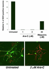
The effect of Ara-C and ribavirin on BDV dissemination and on the subcellular localization of BDV-N in primary hippocampal neurons. The graph on top shows results for primary rat hippocampal neurons at day 7 postinfection (MOI, 0.002) that were doubly stained for BDV-N and the neuronal cell marker MAP-2. Percentages of MAP-2-positive neurons that were also positive for BDV-N were determined. Results are given as percentages of untreated controls. Immunofluorescence images show primary rat hippocampal neurons at day 7 postinfection (MOI, 0.025) doubly stained for BDV-N (green) and MAP-2 (red). Original magnification, ×200.
FIG. 7.
FIG. 7.
Flow cytometric analysis of the effect of treatment with mitotic inhibitors aphidicolin or camptothecin on BDV cell-to-cell spread. Confluent Vero cells were labeled with CFDA and subsequently cocultivated for 5 days with unlabeled, BDV-infected Vero cells (Vero-BV). Cocultivation took place under daily treatment with 4 μM Ara-C, 0.1 mM aphidicolin, or 0.1 μM camptothecin or under treatment every other day with mitotic inhibitors (10 mg of 5-fluoro-2′-deoxyuridine/ml and 25 mg of uridine/ml). The percentages shown indicate the double-positive population within the population of viable cells, which indicates the amount of viral spread. The negative control consisted of a 1:1 mix of CFDA-labeled Vero cells with Vero-BV cells, fixed directly after mixing.
FIG. 8.
FIG. 8.
Ara-C inhibits BDV replication in the brains of rats infected with BDV and prevents the development of persistent CNS infection. (Top) Northern blot analysis. Rats were infected intracranially with BDV and treated with either Ara-C or PBS (mock). At 25 days postinfection rats were sacrificed and total cellular RNA was extracted from brain tissue. Northern blots were probed against cyclophylin (as a housekeeping gene) and BDV-N. The expected sizes of the different BDV mRNAs and genomic RNA are indicated. (Bottom) Histology of brain samples collected from rats infected with BDV and treated with either Ara-C or PBS (mock). Ten-micrometer-thick sections were stained with hematoxylin and eosin and analyzed for the presence of inflammatory infiltrates characteristic for viral persistence in the CNS. Original magnification, ×100.

References

    1. Bautista, J. R., G. J. Schwartz, J. C. de la Torre, T. H. Moran, and K. M. Carbone. 1994. Early and persistent abnormalities in rats with neonatally acquired Borna disease virus infection. Brain Res. Bull. 34:31-36. - PubMed
    1. Bode, L., D. E. Dietrich, R. Stoyloff, H. M. Emrich, and H. Ludwig. 1997. Amantadine and human Borna disease virus in vitro and in vivo in an infected patient with bipolar depression. Lancet 349:178-179. - PubMed
    1. Bode, L., P. Reckwald, W. E. Severus, R. Stoyloff, R. Ferszt, D. E. Dietrich, and H. Ludwig. 2001. Borna disease virus-specific circulating immune complexes, antigenemia, and free antibodies—the key marker triplet determining infection and prevailing in severe mood disorders. Mol. Psychiatry 6:481-491. - PubMed
    1. Bode, L., W. Zimmermann, R. Ferszt, F. Steinbach, and H. Ludwig. 1995. Borna disease virus genome transcribed and expressed in psychiatric patients. Nat. Med. 1:232-236. - PubMed
    1. Carbone, K. M. 2001. Borna disease virus and human disease. Clin. Microbiol. Rev. 14:513-527. - PMC - PubMed

Publication types

MeSH terms

LinkOut - more resources