. 2002 Jun;76(12):6293-301.
doi: 10.1128/jvi.76.12.6293-6301.2002.

Cowpea mosaic virus 32- and 60-kilodalton replication proteins target and change the morphology of endoplasmic reticulum membranes

Affiliations

Cowpea mosaic virus 32- and 60-kilodalton replication proteins target and change the morphology of endoplasmic reticulum membranes

Jan E Carette et al. J Virol. 2002 Jun.

Abstract

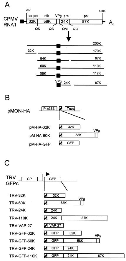
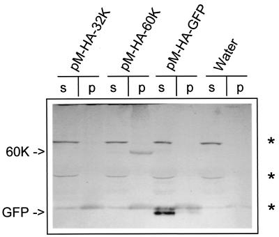
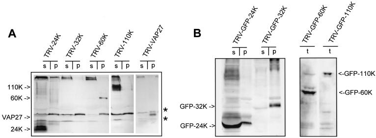
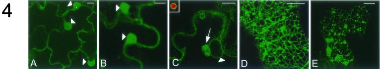
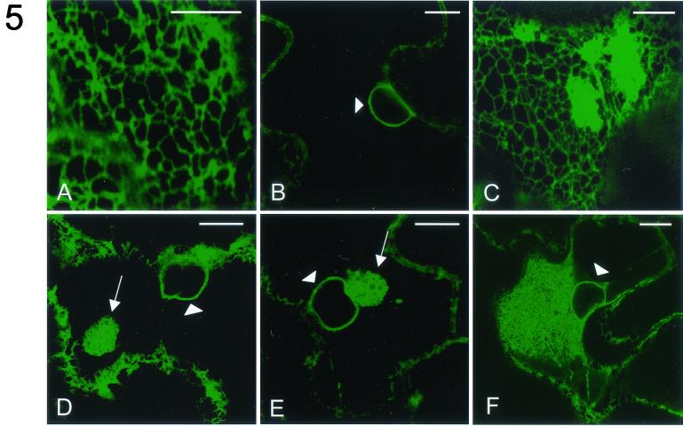
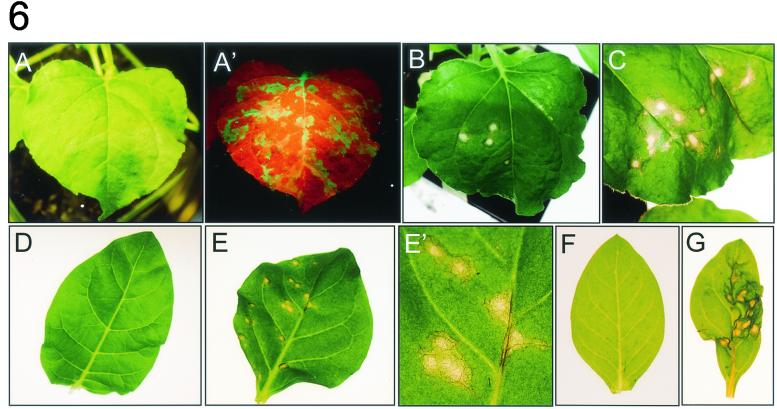
Cowpea mosaic virus (CPMV) replicates in close association with small membranous vesicles that are formed by rearrangements of intracellular membranes. To determine which of the viral proteins are responsible for the rearrangements of membranes and the attachment of the replication complex, we have expressed individual CPMV proteins encoded by RNA1 in cowpea protoplasts by transient expression and in Nicotiana benthamiana plants by using the tobacco rattle virus (TRV) expression vector. The 32-kDa protein (32K) and 60K, when expressed individually, accumulate in only low amounts but are found associated with membranes mainly derived from the endoplasmic reticulum (ER). 24K and 110K are freely soluble and accumulate to high levels. With the TRV vector, expression of 32K and 60K results in rearrangement of ER membranes. Besides, expression of 32K and 60K results in necrosis of the inoculated N. benthamiana leaves, suggesting that 32K and 60K are cytotoxic proteins. On the other hand, during CPMV infection 32K and 60K accumulate to high levels without causing necrosis.


Figures

FIG. 1.
FIG. 1.
Schematic diagram of CPMV RNA1 and the TRV RNA2-based expression vector used to express the CPMV nonstructural proteins. (A) Genetic organization and translational expression of CPMV RNA1. Open reading frames in the RNA molecules (open bars), VPg (black square), cleavage sites (Q/G, Q/M, and Q/S), and nucleotide positions of start and stop codons are indicated. Abbreviations: pro, proteinase; co-pro, cofactor for proteinase; ntb, nucleotide binding protein; pol, core RdRp. (B) Organization of the constructs in the plant expression vector pMON-HA used to transiently express CPMV RNA1-encoded proteins in cowpea protoplasts. Expression is driven by an enhanced CaMV 35S promoter (P-e35S), and transcription is terminated at a nopaline synthase terminator sequence (Tnos). An HA epitope tag was added to facilitate immunodetection of the expressed proteins (hatched box). 60K is composed of the 58K nucleotide-binding protein and VPg. (C) Organization of the expression vector TRV-GFPc and the TRV constructs used to express CPMV RNA1-encoded proteins in N. benthamiana leaves. The duplicated promoter regionwith downstream cloning sites (arrow) permits transcription of a subgenomic RNA for expression of foreign gene inserts. The 110K polymerase is composed of the 24K proteinase and the 87K core polymerase as indicated. Additional constructs were made that contain a fusion with GFP. VAP27 is an A. thaliana protein that is inserted in the ER membrane. The position of the HA epitope tag is indicated with a hatched box.
FIG. 2.
FIG. 2.
Immunodetection of CPMV RNA1-encoded proteins in different subcellular fractions of cowpea protoplasts transfected with transient expression vectors. Protoplasts were harvested 16 h posttransfection, and homogenates were separated in the supernatant (s) and pellet fraction (p) by centrifugation at 30,000 × g. Detection of the proteins was done with a mouse monoclonal anti-HA antibody. 60K and GFP are indicated by arrows. Cowpea proteins that cross-react with this antibody are indicated by asterisks. “Water” indicates lanes with no DNA added.
FIG. 3.
FIG. 3.
Immunodetection of CPMV RNA1-encoded proteins, either tagged with HA (A) or fused to GFP (B), in different subcellular fractions of N. benthamiana leaves infected with TRV expression vectors. Infected leaves were collected 1 dpi, and total homogenates (t) were prepared that were either loaded directly for TRV-GFP-60K and TRV-GFP-110K or centrifuged at 30,000 × g to separate proteins from the supernatant (s) and pellet fraction (p) for the other constructs. Detection of the proteins was done with mouse monoclonal anti-HA antibody (A) or with a rabbit polyclonal anti-GFP serum (B). The bands corresponding to the CPMV proteins are indicated by arrows, while N. benthamiana proteins that cross-react with anti-HA are indicated by asterisks.
FIG.4.
FIG.4.
Subcellular distribution of CPMV proteins fused to GFP in N. benthamiana epidermal cells infected with TRV expression vectors. Fluorescent signals were visualized by confocal microscopy 1 dpi. GFP-110K (A) is present in the nucleus (arrowheads) and in a diffuse pattern in the cytoplasm, as is nonfused GFP (B). GFP-60K (C) localizes in a ring surrounding the nucleus (arrowhead), in one or several bodies (arrow) often near the nucleus, and in a ring surrounding plastids that autofluoresce in red (inset). GFP-32K localizes in a cortical reticulated network (D) and in cortical fluorescent aggregates (E). The images shown in panels A to C correspond to single optical sections of 1 μm, while panels D and E correspond to projections of serial optical sections. Bars, 10 μm.
FIG.5.
FIG.5.
Effect of expression of different CPMV proteins on the morphology of the ER. N. benthamiana plants transgenic with GFP targeted to the lumen of the ER (ER-GFP) were infected with TRV expression vectors. Fluorescent signals were visualized by confocal microscopy 1 dpi. In epidermal cells infected with TRV the pattern of ER-GFP fluorescence is not distinguishable from that in mock-infected cells and is present in a cortical reticulate pattern (A) and in a ring around the nucleus (arrowhead; B). In cells infected with TRV-32K ER-GFP fluorescence is additionally present in large aggregates connected to the cortical network (C) and in one or several spherical aggregates (arrow; D) often near the nucleus (arrowhead). In TRV-60K-infected cells the latter structure is also present (arrow; E). Extensive ER proliferation near the nucleus (arrowhead) of a CPMV-infected cell is shown (F). The images shown in panels B, D, E, and F correspond to single optical sections of 1 μm, while panels A and C correspond to projections of serial optical sections. Bars, 10 μm.
FIG.6.
FIG.6.
Necrosis of leaves from different host plants infected with TRV-32K and TRV-60K. Leaves were infected with the TRV constructs and photographed 3 dpi. (A and A′) N. benthamiana leaf infected with TRV-GFP photographed either under white light (A) or under UV light (A′), where GFP fluorescence appears green. The red fluorescence in A′ is due to chlorophyll. (B to G) Leaves were photographed under white light. (B) N. benthamiana leaf infected with TRV-32K. (C) N. benthamiana leaf infected with TRV-60K. (D) N. tabacum leaf infected with TRV-GFP. (E) N. tabacum leaf infected with TRV-32K. (E′) Close-up of panel E. (F) N. clevelandii infected with TRV-GFP. (G) N. clevelandii infected with TRV-60K.

Similar articles

Cited by

References

    1. Aldabe, R., A. Barco, and L. Carrasco. 1996. Membrane permeabilization by poliovirus proteins 2B and 2BC. J. Biol. Chem. 271:23134-23137. - PubMed
    1. Aldabe, R., and L. Carrasco. 1995. Induction of membrane proliferation by poliovirus proteins 2C and 2BC. Biochem. Biophys. Res. Commun. 206:64-76. - PubMed
    1. Barco, A., and L. Carrasco. 1995. A human virus protein, poliovirus protein 2BC, induces membrane proliferation and blocks the exocytic pathway in the yeast Saccharomyces cerevisiae. EMBO J. 14:3349-3364. - PMC - PubMed
    1. Barco, A., and L. Carrasco. 1998. Identification of regions of poliovirus 2BC protein that are involved in cytotoxicity. J. Virol. 72:3560-3570. - PMC - PubMed
    1. Bertens, P., J. Wellink, R. Goldbach, and A. van Kammen. 2000. Mutational analysis of the cowpea mosaic virus movement protein. Virology 267:199-208. - PubMed

Publication types

LinkOut - more resources