. 2002 Jun;76(12):6311-22.
doi: 10.1128/jvi.76.12.6311-6322.2002.

Early pathogenesis of transmucosal feline immunodeficiency virus infection

Affiliations

Early pathogenesis of transmucosal feline immunodeficiency virus infection

Leslie A Obert et al. J Virol. 2002 Jun.

Abstract

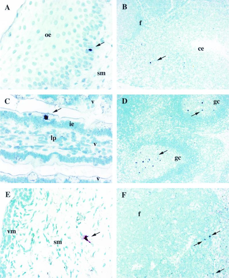
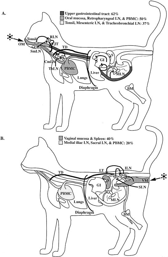
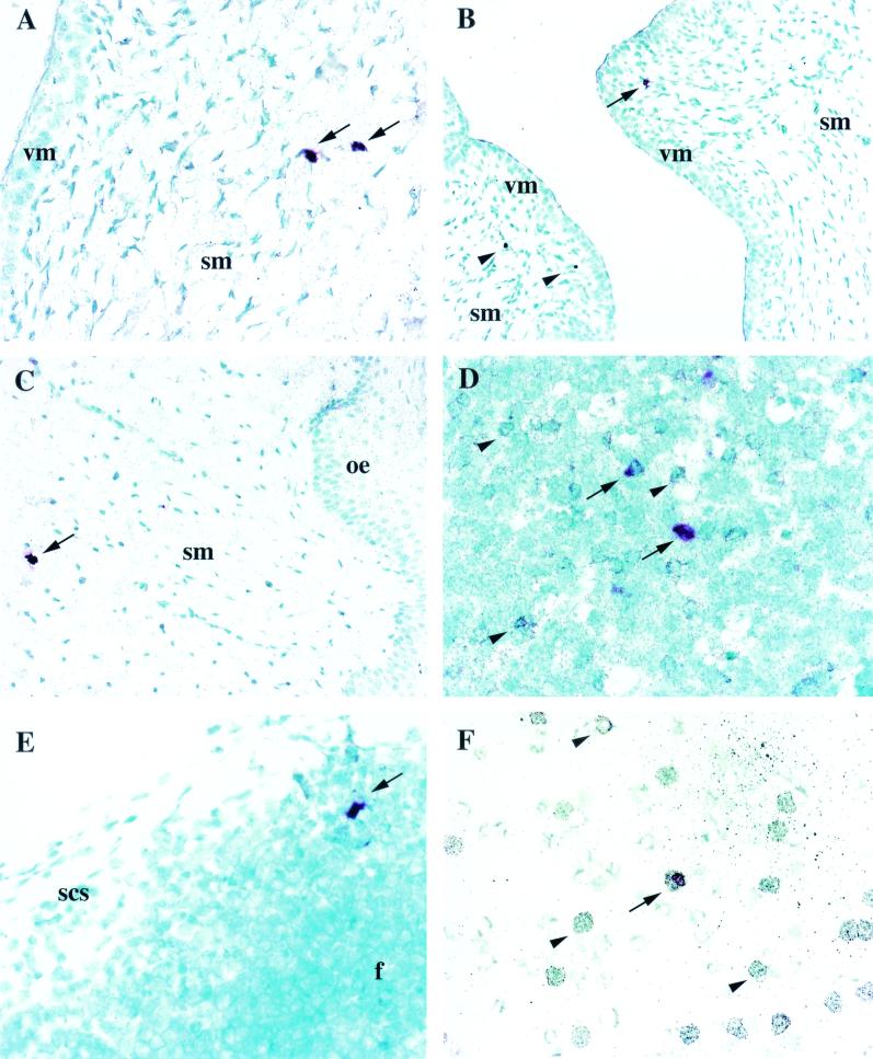
To identify the early target cells and tissues in transmucosal feline immunodeficiency virus (FIV) infection, cats were exposed to a clade C FIV isolate via the oral-nasal or vaginal mucosa and multiple tissues were examined by virus isolation coculture (VI), DNA PCR, catalyzed tyramide signal-amplified in situ hybridization (TSA-ISH), and immunohistochemistry between days 1 and 12 postinoculation (p.i.). FIV RNA was detected in tonsil and oral or vaginal mucosa as early as 1 day p.i. by TSA-ISH and in retropharyngeal, tracheobronchial, or external iliac lymph nodes and sometimes in spleen or blood mononuclear cells by day 2, indicating that regional and distant spread of virus-infected cells occurred rapidly after mucosal exposure. By day 8, viral RNA, DNA, and culturable virus were uniformly detected in regional and distant tissues, connoting systemic infection. TSA-ISH proved more sensitive than DNA PCR in detecting early FIV-infected cells. In mucosal tissues, the earliest demonstrable FIV-bearing cells were either within or subjacent to the mucosal epithelium or were in germinal centers of regional lymph nodes. The FIV(+) cells were of either of two morphological types, large stellate or small round. Those FIV RNA(+) cells which could be colabeled for a phenotype marker, were labeled for either dendritic-cell-associated protein p55 or T-lymphocyte receptor antigen CD3. These studies indicate that FIV crosses mucous membranes within hours after exposure and rapidly traffics via dendritic and T cells to systemic lymphoid tissues, a pathway similar to that thought to occur in the initial phase of infection by the human and simian immunodeficiency viruses.


Figures

FIG. 1.
FIG. 1.
RNA ISH utilizing either Vector red or BCIP-nitroblue tetrazolium chromogen and methyl green counterstain. (A) FIV+ cell (arrow) in the oral epithelium (oe) of cat 3797 1 day p.i. Magnification, ×400. (B) FIV+ cell (arrow) in the tonsillar parenchyma adjacent to the crypt epithelium (ce) of cat 3791 2 days p.i. Magnification, ×100. (C) FIV+ cell (arrow) in the intestinal epithelium (ie) of a villus (v) from the jejunum of cat 3791 2 days p.i. Magnification, ×400. lp, lamina propria. (D) Numerous FIV+ cells (arrows) in the germinal center (gc) of a follicle (f) from the retropharyngeal LN from cat 3795 10 days p.i. Magnification, 100. (E) FIV+ cell (arrow) with DC morphology in the vaginal submucosa (sm) of cat 3598 2 days p.i. Magnification, ×400. vm, vaginal mucosa. (F) Several FIV+ cells (arrows) in the parafollicular region of the medial iliac LN from cat 3853 4 days p.i. Magnification, ×200.
FIG. 2.
FIG. 2.
(A) Illustration demonstrating target tissue infection frequency between 1 and 3 days after oral-nasal FIV exposure as determined by DNA PCR and RNA ISH. FIV was most frequently identified in tissues of the upper gastrointestinal tract (GI) (62% of the animals). The next most frequently observed FIV-infected tissues were oral mucosa (OM), retropharyngeal LN (RLN), and PBMC (50% of the animals). (B) Illustration demonstrating target tissue infection frequency between 1 and 3 days after vaginal FIV exposure as determined by DNA PCR and RNA ISH. FIV was most frequently identified in tissues of the vaginal mucosa (VM) and spleen (S) (40% of the animals). The next most frequently observed FIV-infected tissues were medial iliac LN (ILN), sacral LN (SLN), and PBMC (20% of the animals). Arrows (both panels), flow of lymphatic drainage from the mucosal tissues to regional lymphoid tissues to lymphatic trunks and/or ducts, which ultimately empty into the venous circulation (PBMC). BM, bone marrow; CmLN, cranial mediastinal LN; JT, jugular trunk; LT, lumbar trunk; SmLN, submandibular LN; T, thymus; TbLN, tracheobronchial LN; TD, thoracic duct.
FIG. 3.
FIG. 3.
Dual RNA ISH and immunohistochemistry utilizing Vector red for FIV RNA probe detection, silver-enhanced immunogold for phenotypic identification, and methyl green counterstain. (A) p55+ FIV+ DC (arrows) in the vaginal submucosa (sm) of cat 3826 6 days p.i. Magnification, ×400. vm, vaginal mucosa. (B) FIV+ cell (arrow) and two FIV-uninfected MAC387+ macrophages (arrowheads) in the vaginal submucosa of cat 3826 6 days p.i. Magnification, ×200. (C) p55+ FIV+ cell (arrow) in the oral submucosa of cat 3804 10 days p.i. Magnification, ×200. oe, oral epithelium. (D) CD3+ FIV+ T cells (arrows) and several FIV-uninfected CD3+ cells (arrowheads) in the tonsillar parenchyma of cat 3795 10 days p.i. Magnification, ×400. (E) MAC387+ FIV+ macrophage (arrow) in a follicle (f) adjacent to the subcapsular sinus (scs) region of the retropharyngeal LN from cat 3815 3 days p.i. Magnification, ×400. (F) CD3+ FIV+ T cell (arrow) and several FIV-uninfected CD3+ T cells (arrowheads) in the PBMC of cat 3804 10 days p.i. Magnification, ×400.

Similar articles

Cited by

References

    1. Adachi, A., S. Koenig, H. E. Gendelman, D. Daugherty, S. Gattoni-Celli, A. S. Fauci, and M. A. Martin. 1987. Productive, persistent infection of human colorectal cell lines with human immunodeficiency virus. J. Virol. 61:209-213. - PMC - PubMed
    1. Amerongen, H. M., R. Weltzin, C. M. Farnet, P. Michetti, W. A. Haseltine, and M. R. Neutra. 1991. Transepithelial transport of HIV-1 by intestinal M cells: a mechanism for transmission of AIDS. J. Acquir. Immune Defic. Syndr. 4:760-765. - PubMed
    1. Ayehunie, S., R. W. Groves, A. M. Bruzzese, R. M. Ruprecht, T. S. Kupper, and E. Langhoff. 1995. Acutely infected Langerhans cells are more efficient than T cells in disseminating HIV type 1 to activated T cells following a short cell-cell contact. AIDS Res. Hum. Retrovir. 11:877-884. - PubMed
    1. Baba, T. W., A. M. Trichel, L. An, V. Liska, L. N. Martin, M. Murphey-Corb, and R. M. Ruprecht. 1996. Infection and AIDS in adult macaques after nontraumatic oral exposure to cell-free SIV. Science 272:1486-1489. - PubMed
    1. Bach, J. M., M. Hurtrel, L. Chakrabarti, J. P. Ganiere, L. Montagnier, and B. Hurtrel. 1994. Early stages of feline immunodeficiency virus infection in lymph nodes and spleen. AIDS Res. Hum. Retrovir. 10:1731-1738. - PubMed

Publication types

MeSH terms