. 2002 Jun;76(12):6323-31.
doi: 10.1128/jvi.76.12.6323-6331.2002.

Deubiquitinating function of adenovirus proteinase

Affiliations

Deubiquitinating function of adenovirus proteinase

Maxim Y Balakirev et al. J Virol. 2002 Jun.

Abstract

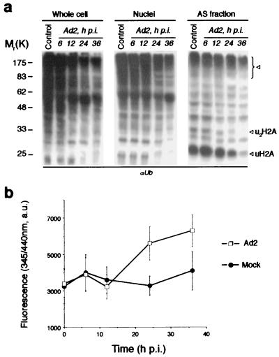
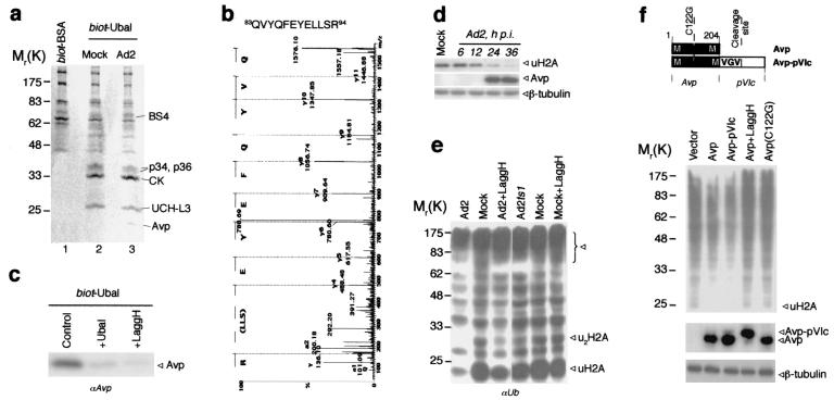
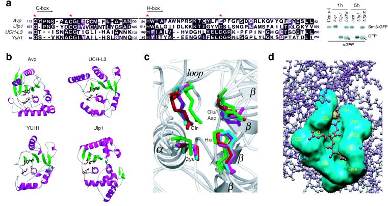
The invasion strategy of many viruses involves the synthesis of viral gene products that mimic the functions of the cellular proteins and thus interfere with the key cellular processes. Here we show that adenovirus infection is accompanied by an increased ubiquitin-cleaving (deubiquitinating) activity in the host cells. Affinity chromatography on ubiquitin aldehyde (Ubal), which was designed to identify the deubiquitinating proteases, revealed the presence of adenovirus L3 23K proteinase (Avp) in the eluate from adenovirus-infected cells. This proteinase is known to be necessary for the processing of viral precursor proteins during virion maturation. We show here that in vivo Avp deubiquitinates a number of cellular proteins. Analysis of the substrate specificity of Avp in vitro demonstrated that the protein deubiquitination by this enzyme could be as efficient as proteolytic processing of viral proteins. The structural model of the Ubal-Avp interaction revealed some similarity between S1-S4 substrate binding sites of Avp and ubiquitin hydrolases. These results may reflect the acquisition of an advantageous property by adenovirus and may indicate the importance of ubiquitin pathways in viral infection.


Figures

FIG. 1.
FIG. 1.
Deubiquitinating activity in Ad-infected cells. (a) Decline of the Ub conjugates pool in Ad2-infected cells. HeLa cells were infected with Ad2 (multiplicity of infection = 100), whereupon cells were harvested at the indicated time p.i. and whole cell, nuclear, and AS nuclear extracts were prepared. The same amount of the protein from each sample was resolved by SDS-PAGE and analyzed by Western blotting with anti-Ub antibodies. Lane control, extracts from mock-infected cells. Arrows indicate the Ub conjugates significantly diminished during Ad2 infection. The uH2A band was also recognized by anti-uH2A MAb E6C5 (not shown). (b) Ad2-infected cells demonstrate increased DUB activity. Whole cell lysates from Ad2- and mock-infected cells were incubated with zLRGG-AMC fluorogenic substrate (100 μM, 1 h) and the fluorescence of liberated AMC was measured at 345 nm/440 nm. Values are means ± standard deviations (error bars) from three separate experiments
FIG. 2.
FIG. 2.
Identification of Ad proteinase Avp as a DUB. (a) The proteins purified by Ubal-affinity chromatography. Whole cell lysates were incubated with biot-BSA or biot-Ubal (20 ng/mg of cell protein, 1 h, 4°C), and loaded on streptavidin agarose column. After extensive washing the bound proteins were eluted with DTT-urea. The eluates from Ad2 (line 1, 3) and mock-infected (line 2) cells were resolved by SDS-PAGE and stained with Coomassie blue. The proteins identified by mass spectrometry are shown. (b) The MS/MS spectrum of Avp-derived peptide obtained from 23K band (Fig. 2a, line 3). (c) Competition of Ubal and LaggH for Avp retrieval on biot-Ubal/streptavidin column. The conditions were as for Fig. 2a, but 10 times less cell lysate was used (control). Ubal (fivefold excess over biot-Ubal) or LaggH (100 μM) was added in the incubation buffer. Western blot from eluates was probed with anti-Avp antiserum. (d) Simultaneous analysis of uH2A and Avp in Ad2-infected cells. Whole cell lysates were prepared at the indicated time p.i., and analyzed by Western blotting with anti-uH2A MAb E6C5 and anti-Avp antiserum (probing with anti-β-tubulin IgG served a control). (e) Contribution of Avp activity to protein deubiquitination during Ad infection. Cells were infected with Ad2, Adts1, or Ad2 in the presence of LaggH (100 μM) for 24 h at nonpermissive temperature (39.5°C). The same amount of AS protein was analyzed by Western blotting with anti-Ub antibody. (f) Deubiquitinating activity of Avp in vivo. HeLa cells were cotransfected with His6-tagged ubiquitin and either control empty vector, or the vector with wild-type proteinase Avp, Avp-pVIc fusion, or inactive mutant Avp(C122G). In Avp-pVIc construct the pVIc activating peptide GVQSLKRRRCF is separated from proteinase sequence by MVGV↓G Avp cleavage site. After 24 h of incubation in the presence or absence of 100 μM LaggH, cells were lysed in 8 M urea, and His6Ub conjugates were isolated by using Ni-NTA resin and analyzed by Western blotting with anti-Ub antibody. The same cell lysates were also analyzed for Avp expression with anti-Avp antiserum (probing with anti-β-tubulin IgG served as a control).
FIG. 3.
FIG. 3.
Substrate specificity of Avp. (a) Cleavage of tetra-Ub and pro-ISG15 by recombinant Avp in the presence of pVIc peptide. Reactions were stopped at different times with NEM (50 mM) and analyzed by Western blotting with rabbit anti-Ub and anti-ISG15 antibodies. Where indicated, Avp was pretreated with the inhibitors LaggH (1 μM) or Ubal (1 μM) for 5 min at 4°C. In one case the activating peptide pVIc was omitted from reaction mixture. (b) Chromatographic analysis of Avp species on unoS column: Avp alone (I), Avp incubated with pVIc for 15 min (II), and Avp incubated with pVIc and Ubal for 30 min (III). (c) Cleavage of Ub-GST and Ub-pVIc-GST fusion proteins by Avp. The reaction conditions were as for Fig. 3a. The Avp cleavage sites are indicated in bold, whereas the pVIc-derived sequence is in italics. The anti-GST MAb B-14 and rabbit anti-Ub antibody were used for Western blotting.
FIG. 4.
FIG. 4.
Structural comparison of Avp with UCH and Ulp families of enzymes. (a) Sequence alignment of the catalytic core domains (Cys and His boxes) of Avp, human UCH-L3, yeast YUH1, and yeast Ulp1 (MEGALIGN [6]). Asterisks indicate the residues of the catalytic triad and Gln residue of the oxyanion hole. Western blot shows the comparative cleavage of Smt3-GFP fusion by Avp, Ulp1, and deubiqutinating enzyme USP8. White arrows show the protein present in crude USP8 preparation cross-reacting with anti-GFP antibody. (b) Ribbon diagrams of the peptidases (PDB identifiers 1AVP, 1UCH, 1CMX, 1EUV). The side chains of the catalytic residues and Gln residue of the oxyanion hole are shown. Figure 4b and c were prepared with MOLSCRIPT (19) and RASTER3D (27). (c) Comparison of the peptidase active sites. Superposition of the active site residues, obtained with the program LSQMAN (CCP4 suite [4]). Avp is shown in magenta, UCH-L3 s shown in green, YUH1 s shown in cyan, and Ulp1 s shown in red. Secondary structure elements of Avp are labeled. (d) Model of the binding of ubiquitin C-terminal peptide LRG-Glz (red) to the active-site cleft of Avp (cyan). The active site tetrads of Avp and YUH1 (in the complex with Ubal) were aligned, and the residues involved in Ubal binding by Avp were identified by determination of the contacts between LRG-Glz peptide and Avp (CCONTACT, CCP4 suite [4]).

References

    1. Anderson, C. W. 1990. The proteinase polypeptide of adenovirus serotype 2 virions. Virology 177:259-272. - PubMed
    1. Andres, G., A. Alejo, C. Simon-Mateo, and M. L. Salas. 2001. African swine fever virus protease, a new viral member of the SUMO-1-specific protease family. J. Biol. Chem. 276:780-787. - PubMed
    1. Bertolaet, B. L., D. J. Clarke, M. Wolff, M. H. Watson, M. Henze, G. Divita, and S. I. Reed. 2001. UBA domains of DNA damage-inducible proteins interact with ubiquitin. Nat. Struct. Biol. 8:417-422. - PubMed
    1. CCP4. 1994. The CCP4 suite: programs for protein crystallography. Acta Cryst. D 50:760-763. - PubMed
    1. Chen, P. H., D. A. Ornelles, and T. Shenk. 1993. The adenovirus L3 23-kilodalton proteinase cleaves the amino-terminal head domain from cytokeratin 18 and disrupts the cytokeratin network of HeLa cells. J. Virol. 67:3507-3514. - PMC - PubMed

Publication types

LinkOut - more resources