. 2002 Jun;76(12):6344-55.
doi: 10.1128/jvi.76.12.6344-6355.2002.

Characterization of a highly pathogenic H5N1 avian influenza A virus isolated from duck meat

Affiliations

Characterization of a highly pathogenic H5N1 avian influenza A virus isolated from duck meat

Terrence M Tumpey et al. J Virol. 2002 Jun.

Abstract

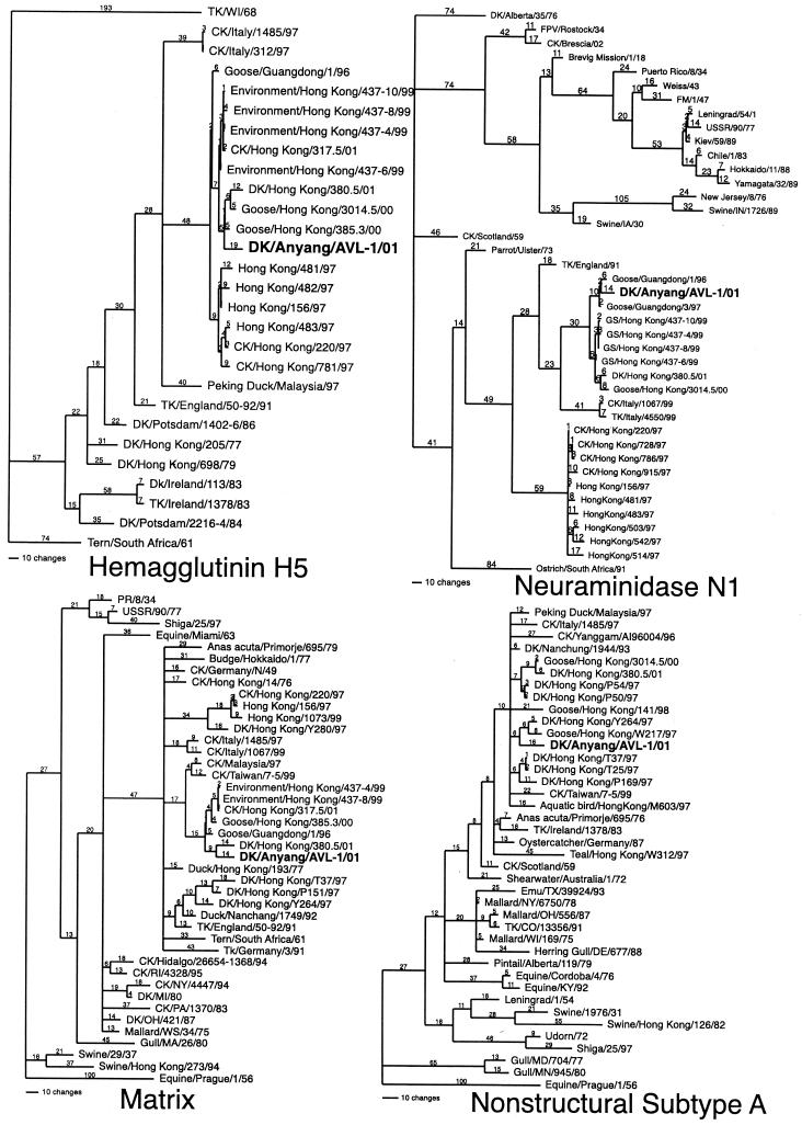
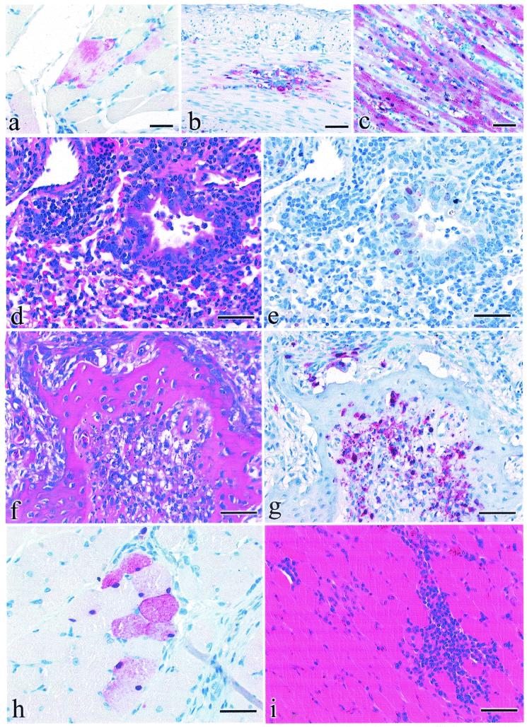
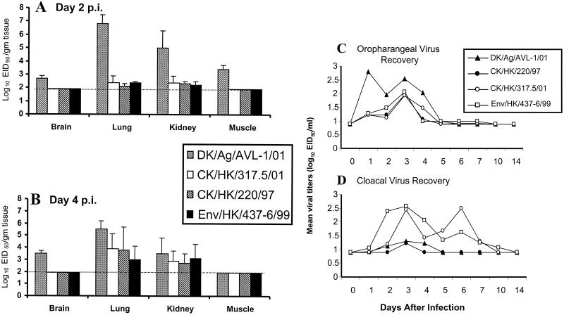
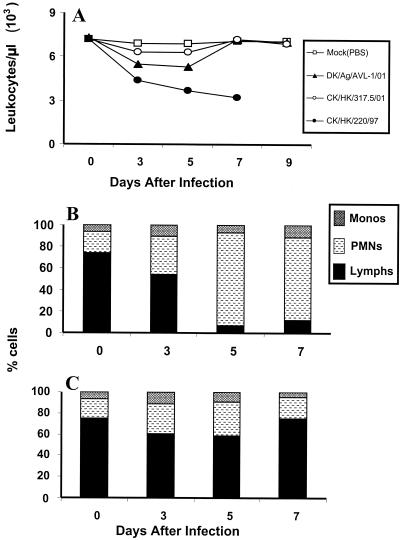
Since the 1997 H5N1 influenza virus outbreak in humans and poultry in Hong Kong, the emergence of closely related viruses in poultry has raised concerns that additional zoonotic transmissions of influenza viruses from poultry to humans may occur. In May 2001, an avian H5N1 influenza A virus was isolated from duck meat that had been imported to South Korea from China. Phylogenetic analysis of the hemagglutinin (HA) gene of A/Duck/Anyang/AVL-1/01 showed that the virus clustered with the H5 Goose/Guandong/1/96 lineage and 1997 Hong Kong human isolates and possessed an HA cleavage site sequence identical to these isolates. Following intravenous or intranasal inoculation, this virus was highly pathogenic and replicated to high titers in chickens. The pathogenesis of DK/Anyang/AVL-1/01 virus in Pekin ducks was further characterized and compared with a recent H5N1 isolate, A/Chicken/Hong Kong/317.5/01, and an H5N1 1997 chicken isolate, A/Chicken/Hong Kong/220/97. Although no clinical signs of disease were observed in H5N1 virus-inoculated ducks, infectious virus could be detected in lung tissue, cloacal, and oropharyngeal swabs. The DK/Anyang/AVL-1/01 virus was unique among the H5N1 isolates in that infectious virus and viral antigen could also be detected in muscle and brain tissue of ducks. The pathogenesis of DK/Anyang/AVL-1/01 virus was characterized in BALB/c mice and compared with the other H5N1 isolates. All viruses replicated in mice, but in contrast to the highly lethal CK/HK/220/97 virus, DK/Anyang/AVL-1/01 and CK/HK/317.5/01 viruses remained localized to the respiratory tract. DK/Anyang/AVL-1/01 virus caused weight loss and resulted in 22 to 33% mortality, whereas CK/HK/317.5/01-infected mice exhibited no morbidity or mortality. The isolation of a highly pathogenic H5N1 influenza virus from poultry indicates that such viruses are still circulating in China and may present a risk for transmission of the virus to humans.


Figures

FIG. 1.
FIG. 1.
Phylogenetic trees of the nucleotide sequences of HA subtype 5, neuraminidase subtype 1, matrix, and nonstructural subtype (group) A, including the isolate DK/Anyang/AVL-1/01 and representative human, swine, equine, and avian influenza virus gene sequences when appropriate. The trees were generated with the PAUP 4 computer program with bootstrap replication (100 bootstraps) and a heuristic search method. The HA tree is rooted to TK/WI/68, the neuraminidase tree is rooted to DK/Alberta/35/76, and the matrix and nonstructural trees are rooted to Equine/Prague/1/56. Branch lengths are included on the tree. Standard two-letter postal codes are used for states in the United States. Abbreviations: TK, turkey; CK, chicken; DK, duck. For isolates without a species, it is assumed to be an isolate from a human.
FIG. 2.
FIG. 2.
Experimental studies of chickens, ducks, and mice inoculated with DK/Anyang/AVL-1/01. Photomicrographs of hematoxylin-and-eosin-stained tissue sections (d, f, and i) or sections stained by IHC methods to demonstrate AIV (a to c, e, g, and h). (a) AIV antigen in cytoplasm of skeletal muscle fibers from a 4-week-old chicken that died 3 days after i.n. inoculation. Bar = 18 μm. (b) AIV antigen in cytoplasm and nuclei of smooth muscle fibers within the tunica muscularis of duodenum from a 4-week-old chicken that died 3 days after i.n. inoculation. Bar = 40 μm. (c) AIV antigen in cytoplasm and nuclei of cardiac muscle fibers from a 4-week-old chicken that died 2 days after i.n. inoculation. Bar = 40 μm. (f) Focal acute periosteal necrosis in pneumatic bone of the cranium in a 2-week-old duck euthanatized 2 days after i.n. inoculation. Bar = 35 μm. (g) AIV antigen in periosteal mesenchymal cells in a 2-week-old duck euthanatized 2 days after i.n. inoculation. Bar = 35 μm. (h) AIV antigen in perilaryngeal skeletal myocytes in a 2-week-old duck euthanatized 2 days after i.n inoculation with DK/Anyang/AVL-1/01 virus. Bar = 15 μm. (i) Focal myofiber degeneration with corresponding lymphohistiocytic myositis in perilaryngeal skeletal muscle in a 2-week-old duck euthanatized 7 days after i.n. inoculation. Bar = 35 μm. (d) Necrotizing bronchitis with neutrophilic inflammation and associated lymphohistiocytic alveolitis in a 4-week-old BALB/c mouse that was euthanatized 4 days after i.n. inoculation. Bar = 35 μm. (e) AIV antigen in nuclei of bronchial epithelium and type II pneumocytes in 4-week-old BALB/c mice that was euthanatized 4 days after i.n. inoculation. Bar = 35 μm.
FIG. 3.
FIG. 3.
Comparison of mean titers of influenza A virus recovered from tissues. Ducks were infected with 106 EID50 of each virus; tissues were collected on days 2 (A) and 4 (B) p.i. and titers in eggs were determined. All isolation attempts without recovery of virus were given a value of 101.9 EID50. This represents the limit of virus detection (horizontal dotted line) for tissues. Mean log10 titers expressed as EID50/milliliter from oropharyngeal (C) and cloacal (D) swabs were sampled from four individual Pekin ducks on the days indicated. For swabs, the limit of virus detection was 100.9 EID50/ml
FIG. 4.
FIG. 4.
Comparison of lung virus titers (A), weight loss (B), and lethality (C) of BALB/c mice infected with 106.0 EID50 of DK/Anyang/AVL-1/01, CK/HK/317.5/01, or CK/HK/220/97 or mock infected. Four to five mice from each virus-infected group were euthanatized on day 4 p.i., and titers in individual lung, trachea, kidney, and brain tissue in embryonated chicken eggs were determined. The limit of virus detection was 101.2 EID50/ml (dotted line). The remaining seven mice from each group were observed for weight loss and mortality through a 14-day observation period.
FIG. 5.
FIG. 5.
Kinetic analysis of circulating leukocytes (A) and blood differential counts (B and C) following H5N1 infection. Two to three mice were infected i.n. with 106.0 EID50 of DK/Anyang/AVL-1/01 (▴), CK/HK/317.5/01 (○), or CK/HK/220/97 (•) or were mock infected (□). Total white blood cell counts were determined by microscopic counting of leukocytes in heparinized whole blood samples diluted with Turks solution. Blood smears were stained with Hema-3 differential stain, and the percentages of monocytes, polymorphonuclear neutrophils (PMNs), and lymphocytes in CK/HK/220/97-infected (B) and DK/Anyang/AVL-1/01-infected (C) mice were determined.

Similar articles

Cited by

References

    1. Alexander, D. J., W. H. Allan, D. G. Parsons, and G. Parsons. 1978. The pathogenicity of four avian influenza viruses for fowls, turkeys and ducks. Res. Vet. Sci. 24:242-247. - PubMed
    1. Alexander, D. J. 1993. Orthomyxovirus infections, p. 287-316. In J. B. McFerran and M. S. McNulty (ed.), Viral infections of vertebrates, vol. III. Viral infections of birds. Elsevier, Amsterdam, The Netherlands.
    1. Alexander, D. J. 2000. A review of avian influenza in different bird species. Vet. Microbiol. 74:3-13. - PubMed
    1. Bankowski, R. A. 1981. Introduction and objectives of the symposium, p. vii-xiv. In R. A. Bankowski (ed.), Proceedings of the 1st International Symposium on Avian Influenza. United States Animal Health Association, Richmond, Va.
    1. Barbeito, M. S., G. Abraham, M. Best, P. Cairns, P. Langevin, W. G. Sterritt, D. Barr, W. Meulepas, J. M. Sanchez-Vizcaino, and M. Saraza. 1995. Recommended biocontainment features for research and diagnostic facilities where animal pathogens are used. First International Veterinary Biosafety Workshop. Rev. Sci. Tech. 14:873-887. - PubMed

Publication types

MeSH terms

Substances