. 2002 Jun;76(12):6356-63.
doi: 10.1128/jvi.76.12.6356-6363.2002.

Half-life of the duck hepatitis B virus covalently closed circular DNA pool in vivo following inhibition of viral replication

Affiliations

Half-life of the duck hepatitis B virus covalently closed circular DNA pool in vivo following inhibition of viral replication

William R Addison et al. J Virol. 2002 Jun.

Abstract

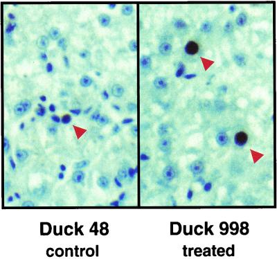
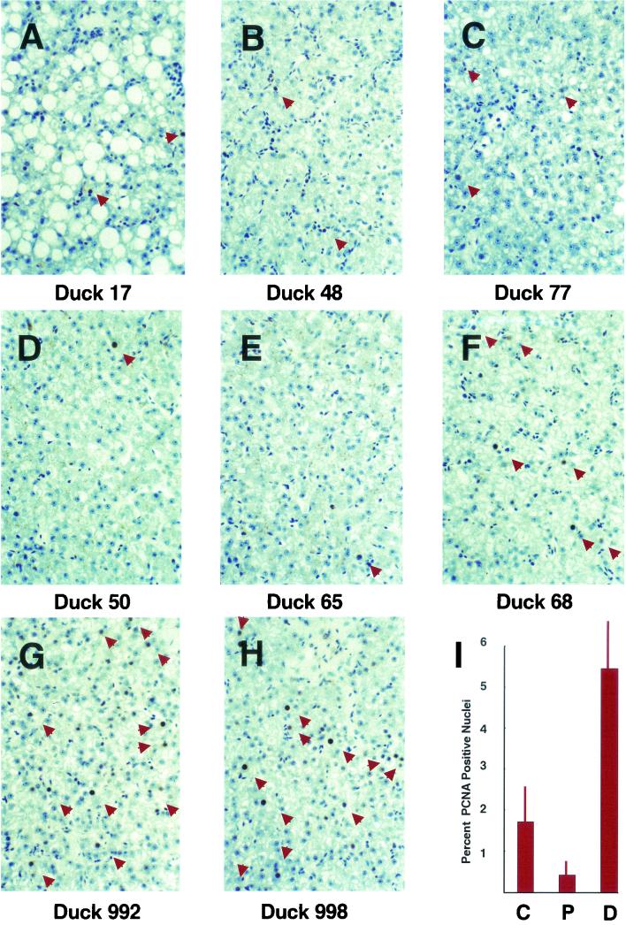
Covalently closed circular DNA (cccDNA) is a crucial intermediate in the replication of hepadnaviruses. We inhibited the replication of duck hepatitis B virus in congenitally infected ducks with a combination of lamivudine and a dideoxyguanosine prodrug. Inhibition of viral replication should prevent renewal of the cccDNA pool, and its decay was measured in liver biopsy samples collected over a 5-month period. In three ducks, the cccDNA pools declined exponentially, with half-lives ranging from 35 to 57 days. In two others, the pools declined exponentially for about 70 days but then stabilized at about 6 copies/diploid genome. The selection of drug-resistant virus mutants is an unlikely explanation for this unexpected stabilization of cccDNA levels. Liver sections stained for the cell division marker PCNA showed that animals in which cccDNA loss was continuous had significantly greater numbers of PCNA-positive nuclei than did those animals in which cccDNA levels had plateaued.


Figures

FIG. 1.
FIG. 1.
(A to C) Serum DHBV DNA levels decline rapidly in ducks treated with lamivudine and a ddG prodrug. The drug treatment and test bleeds of all ducks began at 6 a.m., and antiviral drugs were administered by intramuscular injection every 12 h thereafter. The ducks were initially bled every 6 h, then every 8 h, and finally every 12 h as indicated by the symbols on the graphs. Serum virus levels were measured by dot blot hybridization to a 32P-labeled DHBV probe. Radioactivity was measured with a Fuji phosphorimager, and viremia is reported in arbitrary units (PI units). The birds were housed in a room with a cycle of 10 h of light (commencing at 8 a.m.) followed by 14 h of darkness. (A) Duck 33 was treated with lamivudine and ddG prodrug, and its serum virus level was monitored over 3.5 days. (B) Duck 19 was treated identically to duck 33. (C) Four control ducks (⧫, duck 6; ▪, duck 26; ▴, duck 42; ✖, duck 979) were bled on the same schedule as ducks 33 and 19 but received no drug treatment. (D) Dot blot assay of viremia in ducks. Ten microliters of serum from each duck in the cccDNA half-life study was obtained at each of the indicated time points, applied to a Hybond-N membrane (Amersham), hybridized to a 32P-labeled DHBV probe, and autoradiographed. Ducks 17, 48, and 77 were left untreated, and ducks 50, 65, 68, 992, and 998 were treated with lamivudine and a ddG prodrug. (E) DNA was isolated from serum samples from treated duck 992 at the indicated time points, and DHBV sequences were amplified by PCR. The amount of amplicon for each time point was estimated by measuring the intensity of its band on a stained agarose gel. The amounts of amplicon produced by the indicated serial dilutions of DNA isolated on day 0 are marked by horizontal lines. (F) Quantitative Southern blot of cccDNA present in liver DNA at the start of the half-life study. One microgram of DNA from duck liver tissue (Duck) or uninfected duck erythrocyte doped with known amounts of a plasmid carrying the DHBV genome (Copies DHBV × 106) was digested with EcoRI endonuclease, separated on an agarose gel, transferred to a Hybond-N+ membrane, and probed with 32P-labeled DHBV DNA. A Fuji phosphorimager was used to quantify the hybridized probe. The cccDNA copy numbers inferred from the Southern blot are shown and are compared to the copy numbers derived from the competitive PCR assay.
FIG. 2.
FIG. 2.
DHBV cccDNA has a half-life of 35 to 57 days in ducks treated with lamivudine and ddG prodrug. Drug treatment was performed as described in the legend to Fig. 1. Control ducks were injected with a placebo of phosphate-buffered saline twice per day. At the indicated time points, liver tissue was sampled by needle biopsy, total cellular DNA was isolated, and the cccDNA copy number was measured by competitive PCR. Data from day 43 were anomalous and are shown but were not used in the construction of the curves (see the text). (A) cccDNA copy numbers in untreated ducks (◊, duck 17; □, duck 48; ▵, duck 77); (B to F) decline in cccDNA copy numbers in ducks treated with antiviral drugs.
FIG. 3.
FIG. 3.
Comparison of PCNA staining in control and treated ducks. Liver tissue sections from control duck 48 and treated duck 998 recovered on day 155 of the study were immunostained for the presence of PCNA. PCNA-positive nuclei are indicated by arrowheads. Magnification, ×400.
FIG. 4.
FIG. 4.
Immunohistochemical staining of duck liver sections with anti-PCNA monoclonal antibodies. Liver samples recovered on day 155 of the study were fixed, sectioned, and stained for the presence of PCNA. PCNA-positive nuclei are marked by arrowheads. Sections from control ducks (A to C), ducks in which cccDNA levels plateaued after an initial decline (D and E), and ducks in which cccDNA levels declined through the course of the study (F to H) are shown. The average frequency of PCNA-positive hepatocyte nuclei in three random fields of sections from each duck in each of the three groups (C, control; P, cccDNA plateaud; D, cccDNA declined) is also shown (I). Magnifications, ×160 (all panels)

References

    1. Addison, W. R., W. W. S. Wong, K. P. Fischer, and D. L. J. Tyrrell. 2000. A quantitative competitive PCR assay for the covalently closed circular form of the duck hepatitis B virus. Antivir. Res. 48:27-37. - PubMed
    1. Chyama, K., Y. Suzuki, M. Kobayashi, M. Kobayashi, A. Tsubota, M. Hashimoto, Y. Miyano, H. Koike, M. Kobayashi, I. Koida, Y. Arase, S. Saitoh, N. Murashima, K. Ikeda, and H. Kumada. 1998. Emergence and takeover of YMDD motif mutant hepatitis B virus during long-term lamivudine therapy and re-takeover by wild type after cessation of therapy. Hepatology 27:1711-1716. - PubMed
    1. Civitico, G. M., and S. A. Locarnini. 1994. The half-life of duck hepatitis B virus supercoiled DNA in congenitally infected primary hepatocyte cultures. Virology 203:81-89. - PubMed
    1. Dandri, M., M. R. Burda, H. Will, and J. Petersen. 2000. Increased hepatocyte turnover and inhibition of woodchuck hepatitis B virus replication by adefovir in vitro do not lead to reduction of the closed circular DNA. Hepatology 32:139-146. - PubMed
    1. Dienstag, J. L., R. P. Perrillo, E. R. Schiff, M. Bartholomew, C. Vicary, and M. Rubin. 1995. A preliminary trial of lamivudine for chronic hepatitis B infection. N. Engl. J. Med. 333:1657-1661. - PubMed

Publication types

Associated data

LinkOut - more resources