. 2002 Jun;11(6):1300-8.
doi: 10.1110/ps.4670102.

Epitope mapping of a monoclonal antibody against human thrombin by H/D-exchange mass spectrometry reveals selection of a diverse sequence in a highly conserved protein

Affiliations

Epitope mapping of a monoclonal antibody against human thrombin by H/D-exchange mass spectrometry reveals selection of a diverse sequence in a highly conserved protein

Abel Baerga-Ortiz et al. Protein Sci. 2002 Jun.

Abstract

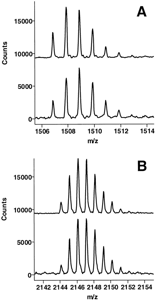
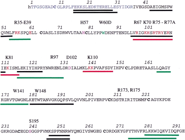
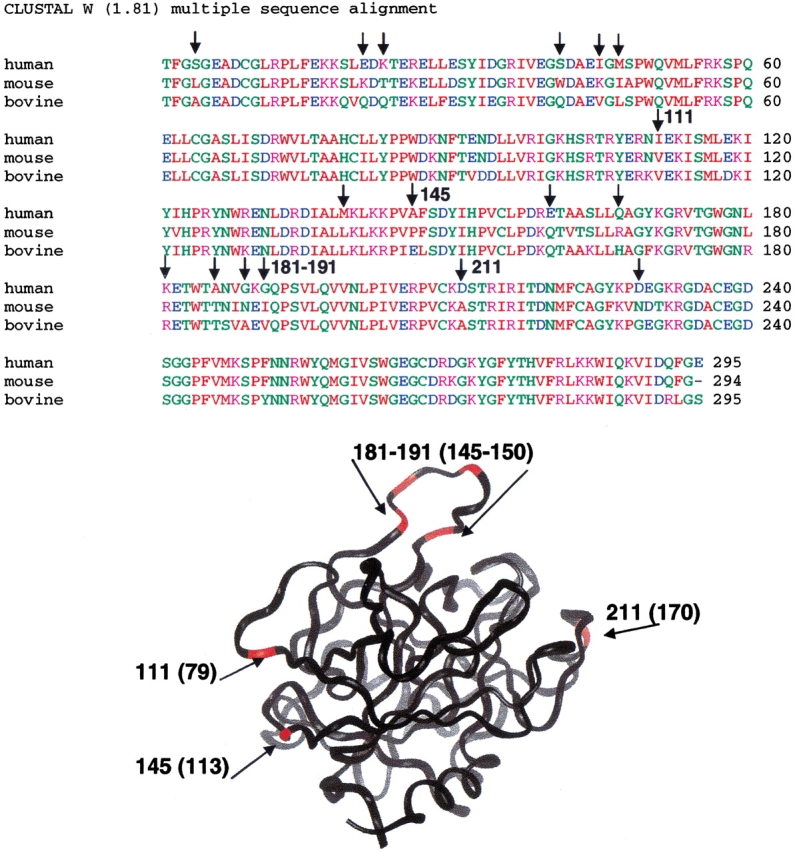
The epitope of a monoclonal antibody raised against human thrombin has been determined by hydrogen/deuterium exchange coupled to MALDI mass spectrometry. The antibody epitope was identified as the surface of thrombin that retained deuterium in the presence of the monoclonal antibody compared to control experiments in its absence. Covalent attachment of the antibody to protein G beads and efficient elution of the antigen after deuterium exchange afforded the analysis of all possible epitopes in a single MALDI mass spectrum. The epitope, which was discontinuous, consisting of two peptides close to anion-binding exosite I, was readily identified. The epitope overlapped with, but was not identical to, the thrombomodulin binding site, consistent with inhibition studies. The antibody bound specifically to human thrombin and not to murine or bovine thrombin, although these proteins share 86% identity with the human protein. Interestingly, the epitope turned out to be the more structured of two surface regions in which higher sequence variation between the three species is seen.


Figures

Fig. 1.
Fig. 1.
The activity of the thrombin:TMEGF456 complex toward protein C was measured after incubating the thrombin with increasing concentrations of the mAb for 10 min (black bars). The mAb had no effect on the TMEGF456-independent activation of protein C (gray bars).
Fig. 2.
Fig. 2.
Binding of thrombin to a TMEGF456 surface was monitored in real time using a BIACORE assay. Sensorgrams were collected both in the presence (black) and in the absence (gray) of mAb. Binding of mAb to thrombin completely abolished the latter's ability to bind to TMEGF456.
Fig. 3.
Fig. 3.
Several regions of thrombin that showed retention of deuterium upon binding of thrombomodulin (Mandell et al. 2001) did not show any significant mass difference upon binding of mAb (bottom spectrum) compared to the no-mAb control (top spectrum). (A) Thrombin residues 167–180 (residues 133–144 in chymotrypsin numbering). (B) Thrombin residues 117–132 (residues 85–101 in chymotrypsin numbering).
Fig. 4.
Fig. 4.
Sequence of thrombin showing the different peptides that were obtained from pepsin digestion and for which quantitative hydrogen/deuterium (H/D) exchange data could be obtained. Key residues are identified above the sequentially numbered sequence in the chymotrypsin numbering scheme. The black bars denote peptides that did not shift in the presence of either TMEGF45 or mAb. The green bars correspond to regions that shifted in mass in the presence of TMEGF45 but not in the presence of the mAb. The red bars show the regions that shifted in mass in the presence of both TMEGF45 and the mAb.
Fig. 5.
Fig. 5.
The region of thrombin corresponding to amino acids 97–117 (residues 67–85 in the chymotrypsin numbering) retained a significant amount of amide deuteration in the presence of the mAb (bottom spectrum) relative to the control (top spectrum).
Fig. 6.
Fig. 6.
The other region of thrombin that was significantly protected in the presence of the mAb was the one corresponding to amino acids 139–149 (residues 108–116 in chymotrypsin numbering). This region also showed some protection upon contact with TMEGF45 (Mandell et al. 2001), but the amount of protection was less.
Fig. 7.
Fig. 7.
Comparison of the binding maps generated by hydrogen/deuterium (H/D) exchange experiments for the interaction of thrombin with mAb (A) and TMEGF45 (B; Mandell et al. 2001). Surface segments of thrombin that were highly protected (blue) are distinguished from those that were only slightly protected (cyan) upon binding of the mAb. Similarly, segments of thrombin that were highly protected (red) are distinguished from those that were only slightly protected (magenta) upon binding of TMEGF45. The binding sites of MAb and TMEGF45 are overlapping but not identical. The thrombin structures are shown in the same orientation and the active site residues are indicated in green.
Fig. 8.
Fig. 8.
Binding of the mAb (10 nM) to a surface containing biotinylated thrombin was monitored in real time both by itself (red) or in the presence of 20 mM (blue) and of 100 mM (green) of the peptide corresponding to residues 97–117 of thrombin (A) and residues 139–149 of thrombin (B). Although the peptide corresponding to residues 97–117 did not affect binding of mAb, the peptide corresponding to residues 139–149 disrupted binding partially at high concentration.
Fig. 9.
Fig. 9.
Alignment of the sequences for human, bovine, and mouse thrombin shows possible sites for antibody recognition. Of 17 sequence differences (vertical arrows above the sequence) between human and mouse thrombin, 8 were on the surface of thrombin and were viable epitope candidates (red in the structure). The residue numbers for these are shown on both the sequence and the structure and they map to different regions of thrombin. Two of them, 111 and 145, were in the identified epitope (arrows), whereas 4 were located near the 181–196 loop, which did not retain deuterium upon binding of mAb.

References

    1. Baerga-Ortiz, A., Rezaie, A.R., and Komives, E.A. 2000. Electrostatic dependence of the thrombin–thrombomodulin interaction. J. Mol. Biol. 296 651–658. - PubMed
    1. Burritt, J., DeLeo, F., McDonald, C., Prigge, J., Dinauer, M., Nakamura, M., Nauseef, W., and Jesaitis, A. 2001. Phage display epitope mapping of human neutrophil flavocytochrome b558. J. Biol. Chem. 276 2053–2061. - PubMed
    1. Chambers, G., Lawrie, L., Cash, P., and Murray, G. 2000. Proteomics: A new approach to the study of disease. J. Pathol. 192 280–288. - PubMed
    1. Dawes, J., James, K., Micklem, L., Pepper, D., and Prowse, C. 1984. Monoclonal antibodies directed against human α-thrombin and the thrombin–antithrombin III complex. Throm. Res. 36 397–409. - PubMed
    1. Fiedler, W., Borchers, C., Macht, M., Deininger, S., and Przybylski, M. 1998. Molecular characterization of a conformational epitope of hen egg white lysozyme by differential chemical modification of immune complexes and mass spectrometric peptide mapping. Bioconjug. Chem. 9 236–241. - PubMed

Publication types

LinkOut - more resources