. 2002 Jun;11(6):1393-408.
doi: 10.1110/ps.2830102.

Protein-protein docking with multiple residue conformations and residue substitutions

Affiliations

Protein-protein docking with multiple residue conformations and residue substitutions

David M Lorber et al. Protein Sci. 2002 Jun.

Abstract

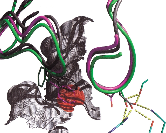
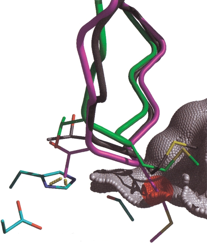
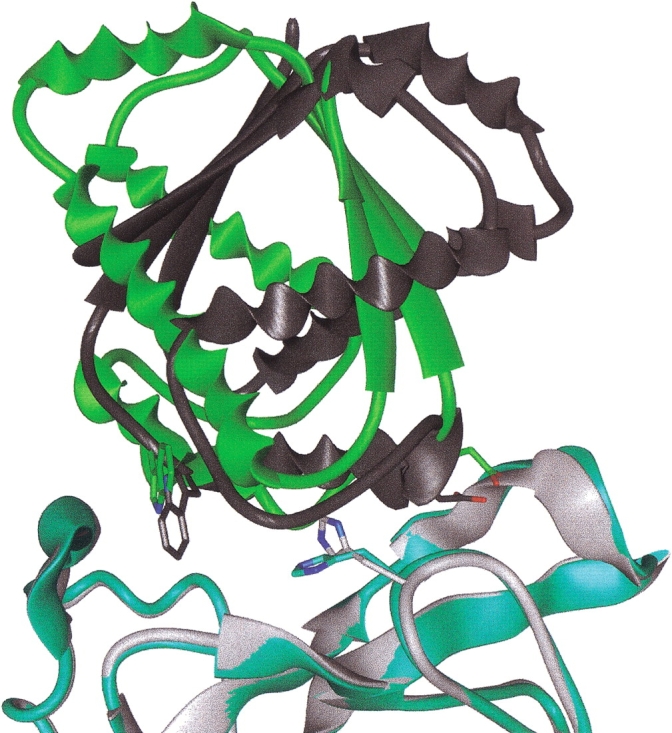
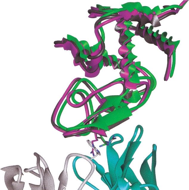
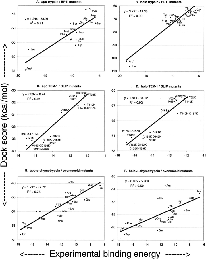
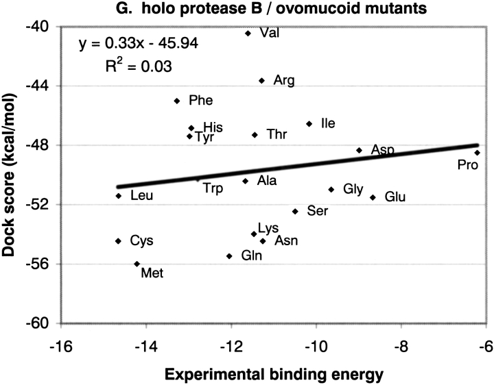
The protein docking problem has two major aspects: sampling conformations and orientations, and scoring them for fit. To investigate the extent to which the protein docking problem may be attributed to the sampling of ligand side-chain conformations, multiple conformations of multiple residues were calculated for the uncomplexed (unbound) structures of protein ligands. These ligand conformations were docked into both the complexed (bound) and unbound conformations of the cognate receptors, and their energies were evaluated using an atomistic potential function. The following questions were considered: (1) does the ensemble of precalculated ligand conformations contain a structure similar to the bound form of the ligand? (2) Can the large number of conformations that are calculated be efficiently docked into the receptors? (3) Can near-native complexes be distinguished from non-native complexes? Results from seven test systems suggest that the precalculated ensembles do include side-chain conformations similar to those adopted in the experimental complexes. By assuming additivity among the side chains, the ensemble can be docked in less than 12 h on a desktop computer. These multiconformer dockings produce near-native complexes and also non-native complexes. When docked against the bound conformations of the receptors, the near-native complexes of the unbound ligand were always distinguishable from the non-native complexes. When docked against the unbound conformations of the receptors, the near-native dockings could usually, but not always, be distinguished from the non-native complexes. In every case, docking the unbound ligands with flexible side chains led to better energies and a better distinction between near-native and non-native fits. An extension of this algorithm allowed for docking multiple residue substitutions (mutants) in addition to multiple conformations. The rankings of the docked mutant proteins correlated with experimental binding affinities. These results suggest that sampling multiple residue conformations and residue substitutions of the unbound ligand contributes to, but does not fully provide, a solution to the protein docking problem. Conformational sampling allows a classical atomistic scoring function to be used; such a function may contribute to better selectivity between near-native and non-native complexes. Allowing for receptor flexibility may further extend these results.


Figures

Fig. 1.
Fig. 1.
Comparing rigid (A, B) and flexible (C, D) docking of an unbound structure of BPTI (4PTI) to unbound (2PTN) and bound (2PTC) trypsin. Data points for the best docking energy scores (y-axis) for all RMSD values (x-axis) are shown. RMSD values are calculated between all Cα atoms of each docked ligand and the Cα atoms of the unbound ligand superpositioned onto the bound ligand. As a convenience to the reader, a black line is drawn to define an envelope of the best energy values. This line includes the lowest RMS data point and 50 additional data points representing the lowest energies in each of 50 divisions of the data. Points for the line are distributed on the x-axis based on the density of the data points (higher resolution where more data points exist). The vertical line at 3 Å indicates an upper RMSD bound for a near-native conformation.
Fig. 2.
Fig. 2.
The high scoring conformation of unbound BPTI (backbone in green) docked in multiple conformations into the unbound conformation of trypsin (surface in gray). The rigid unbound BPTI (magenta) has been superpostioned onto the crystallographic bound BPTI (gray). The molecular surface is colored red where the unbound, rigid BPTI would clash into the surface. The blue surface indicates the position of the catalytic Oγ of Ser195. Asp189 of trypsin and a bridging water molecule (2.6 Å from the docked ligand) are shown for context. No water molecules were present in the docking calculation.
Fig. 3.
Fig. 3.
Lines representing the best docking energy scores (y-axis) versus RMSD from the experimental complex (x-axis) for: (A) TEM-1/BLIP; (B) subtilisin/CI-2; (C) barnase/barstar; (D) FAB/lysozyme; (E) α-chymotrypsin/ovomucoid; (F) protease B/ovomucoid. The dashed lines represent docking the rigid unbound ligand, and the solid lines represent docking the flexible unbound ligand. The blue lines represent docking to the unbound conformations of the receptors, and the pink lines represent docking to the bound conformations of the receptors. A vertical line is drawn at 3 Å to indicate an upper RMSD bound for a near-native conformation.
Fig. 4.
Fig. 4.
Subtraction of the best near-native (RMSD values <3 Å from the bound complex) energy score from the best non-native (>3 Å) energy score for each system. Bars to the left indicate a near-native complex was preferred, and bars to the right indicate that a non-native complex scored best. The magnitude of the bar indicates how well the preferred docked complex is distinguished from other complexes.
Fig. 5.
Fig. 5.
The high-scoring unbound BLIP structure (green), generated from multiconformer docking to the unbound conformation of TEM-1 (cyan), is shown. The rigid unbound BLIP (magenta) has been superimposed onto the bound ligand (gray). A partial molecular surface for the complexed receptor is shown to illustrate hydrophobic interactions. Important intermolecular hydrogen bonds are shown in yellow. The conformations of two key interface residues from BLIP, Asp49 and Phe142, are shown for the best scoring docked structure (green), for the original unbound structure (magenta), and for the bound complex (gray). The molecular surface of TEM-1 is colored red where the superpositioned rigid unbound ligand clashes into the receptor.
Fig. 6.
Fig. 6.
The best-scoring CI-2 ligand (green) generated from multiconformer docking is shown with the unbound conformation of subtilisin (catalytic triad shown in cyan). The rigid unbound CI-2 (magenta) has been superpositioned onto the complexed CI-2 (gray). The dashed red lines indicate clashes between Thr58 of the rigid unbound ligand and the receptor. The red surface indicates the region where the P1 residue from the rigid unbound CI-2 clashes into the receptor surface.
Fig. 7.
Fig. 7.
The best-scoring structure generated from docking multiple conformations of unbound barstar (green) into the unbound conformation of barnase (cyan) is shown. The experimental complex of barstar (gray) and barnase (light gray) has been superimposed on the unbound receptor. The different conformations adopted by His102 of barnase are shown. Glu46 from the unbound structure (green) and Asp36 from the bound structure (gray) are proximal to each other, as are Trp38 (green) and Trp45 (gray).
Fig. 8.
Fig. 8.
The high-scoring conformation of lysozyme from multiconformer docking (green) to the bound structure of FAB D44.1 is shown. The bound form of lysozyme (gray) and the unbound structure of FAB (A, monomer in light gray, B, in cyan) are shown. Arg45 from the unbound ligand structure (magenta), when superpositioned onto the complexed structure of lysozyme, clashes with Trp94 of FAB.
Fig. 9.
Fig. 9.
Comparisons of experimentally determined and docking-predicted binding affinities for mutant protein inhibitors docked into their cognate enzymes. BPTI variants docked to unbound (A) and bound (B) trypsin; BLIP mutants docked to unbound (C) and bound (D) TEM-1; ovomucoid variants docked to unbound (E) and bound (F) α-chymotrypsin, and ovomucoid variants docked to bound protease B (G).
Fig. 9.
Fig. 9.
Comparisons of experimentally determined and docking-predicted binding affinities for mutant protein inhibitors docked into their cognate enzymes. BPTI variants docked to unbound (A) and bound (B) trypsin; BLIP mutants docked to unbound (C) and bound (D) TEM-1; ovomucoid variants docked to unbound (E) and bound (F) α-chymotrypsin, and ovomucoid variants docked to bound protease B (G).

Similar articles

Cited by

References

    1. Buckle, A.M., Schreiber, G., and Fersht, A.R. 1994. Protein–protein recognition: Crystal structural analysis of a barnase–barstar complex at 2.0-A resolution. Biochemistry 33 8878–8889. - PubMed
    1. Camacho, C.J., Gatchell, D.W., Kimura, S.R., and Vajda, S. 2000. Scoring docked conformations generated by rigid-body protein–protein docking. Proteins 40 525–537. - PubMed
    1. Cherfils, J., Duquerroy, S., and Janin, J. 1991. Protein–protein recognition analyzed by docking simulation. Proteins 11 271–280. - PubMed
    1. Claussen, H., Buning, C., Rarey, M., and Lengauer, T. 2001. FlexE: Efficient molecular docking considering protein structure variations. J. Mol. Biol. 308 377–395. - PubMed
    1. Connolly, M.L. 1986. Shape complementarity at the hemoglobin alpha 1 beta 1 subunit interface. Biopolymers 25 1229–1247. - PubMed

Publication types

LinkOut - more resources