. 2002 Jun;11(6):1415-23.
doi: 10.1110/ps.4890102.

An improved hydrogen bond potential: impact on medium resolution protein structures

Affiliations

An improved hydrogen bond potential: impact on medium resolution protein structures

Felcy Fabiola et al. Protein Sci. 2002 Jun.

Abstract

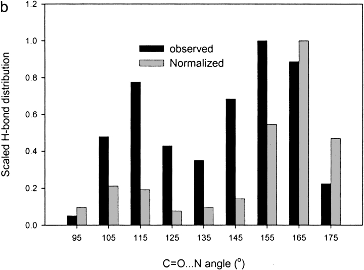
A new semi-empirical force field has been developed to describe hydrogen-bonding interactions with a directional component. The hydrogen bond potential supports two alternative target angles, motivated by the observation that carbonyl hydrogen bond acceptor angles have a bimodal distribution. It has been implemented as a module for a macromolecular refinement package to be combined with other force field terms in the stereochemically restrained refinement of macromolecules. The parameters for the hydrogen bond potential were optimized to best fit crystallographic data from a number of protein structures. Refinement of medium-resolution structures with this additional restraint leads to improved structure, reducing both the free R-factor and over-fitting. However, the improvement is seen only when stringent hydrogen bond selection criteria are used. These findings highlight common misconceptions about hydrogen bonding in proteins, and provide explanations for why the explicit hydrogen bonding terms of some popular force field sets are often best switched off.


Figures

Fig. 1.
Fig. 1.
Main-chain hydrogen bond distances and angles are improved by refinement with the double-well hydrogen bond restraint. A region of chloromucanate cycloisomerase before refinement (a) and after refinement (b) with ɛ = 100. The atoms involved in hydrogen bonding interactions are shown in dark color. N . . . O distances and C = O . . . N angles are indicated.
Fig. 2.
Fig. 2.
Free R-factor (Rfree) versus weight (ɛ) for the main-chain hydrogen bond restraint. Each refinement was performed with optimized hydrogen bond parameters. Optimal ɛ are indicated by arrows.
Fig. 3.
Fig. 3.
Results of refinements without the hydrogen bond restraint, or with the restraint and various parameter sets. The consensus parameters were ɛ = 100, θlow = 115°, θhigh = 155°, Rcut = 3.5 Å, θcut = 90°, and R0 = 2.9 Å.
Fig. 4.
Fig. 4.
Free R-factor (Rfree) versus hydrogen-bond weight for chloromucanate cycloisomerase. Selection of hydrogen bonds is based on two or four criteria. Each refinement was performed with other hydrogen bond parameters fixed at consensus values.
Fig. 5.
Fig. 5.
Normalized distribution of C=O . . . N acceptor angles before inclusion of hydrogen bond restraints in β-catenin (a; at 2.9Å resolution) and arginine kinase (b; at 1.2 Å resolution) and CD2 (c; at 3.1 Å resolution). The gray bars show the distribution normalized by the nonuniform random distribution (Bowie 1997).
Fig. 5.
Fig. 5.
Normalized distribution of C=O . . . N acceptor angles before inclusion of hydrogen bond restraints in β-catenin (a; at 2.9Å resolution) and arginine kinase (b; at 1.2 Å resolution) and CD2 (c; at 3.1 Å resolution). The gray bars show the distribution normalized by the nonuniform random distribution (Bowie 1997).
Fig. 6.
Fig. 6.
Quality of refinements of chloromucanate cycloisomerase using double-well, single-well, and angle-independent hydrogen bond restraints. Consensus restraint parameters were used.
Fig. 7.
Fig. 7.
Distribution of C = O . . . N angles before and after refinement of chloromucanate cycloisomerase with the double-well hydrogen bond restraint (ɛ = 100)
Fig. 8.
Fig. 8.
Refinement of three proteins as a function of the weight applied to side-chain hydrogen bond restraints. A weight of ɛside = 0 implies that no side-chain restraints were applied. Main-chain restraints were included in each case, with the consensus main-chain hydrogen bond parameters.
Fig. 9.
Fig. 9.
Hydrogen bonding restraining potential (EHB). (a) The radial component of the hydrogen bond potential with (EHB, solid) and without (dotted) the switching function (SW). (b) The double-well angular component, for which θmid is the midpoint between θlow and θhigh.
Fig. 9.
Fig. 9.
Hydrogen bonding restraining potential (EHB). (a) The radial component of the hydrogen bond potential with (EHB, solid) and without (dotted) the switching function (SW). (b) The double-well angular component, for which θmid is the midpoint between θlow and θhigh.

Similar articles

Cited by

References

    1. Avila-Sakar, A.J., Creutz, C.E., and Kretsinger, R.H. 1998. Crystal structure of bovine annexin VI in a calcium-bound state. Biochim. Biophys. Acta 1387 103–116. - PubMed
    1. Baker, E.N. and Hubbard, R.E. 1984. Hydrogen bonding in globular proteins. Prog. Biophys. Mol. Biol. 44 97–179. - PubMed
    1. Berman, H.M., Westbrook, J., Feng, Z., Gilliland, G., Bhat, T.N., Weissig, H., Shindyalov, I.N., and Bourne, P.E. 2000. The Protein Data Bank. Nucleic Acids Res. 28 235–242. - PMC - PubMed
    1. Bordo, D. and Argos, P. 1994. The role of side-chain hydrogen bonds in the formation and stabilization of secondary structure in soluble proteins. J. Mol. Biol. 243 504–519. - PubMed
    1. Bowie, J.U. 1997. Helix packing angle preferences. Nat. Struct. Biol. 4 915–917. - PubMed

Publication types

LinkOut - more resources