. 2002 Jun;11(6):1472-81.
doi: 10.1110/ps.0202502.

Site-directed spin labeling of a bacterial chemoreceptor reveals a dynamic, loosely packed transmembrane domain

Affiliations

Site-directed spin labeling of a bacterial chemoreceptor reveals a dynamic, loosely packed transmembrane domain

Alexander Barnakov et al. Protein Sci. 2002 Jun.

Abstract

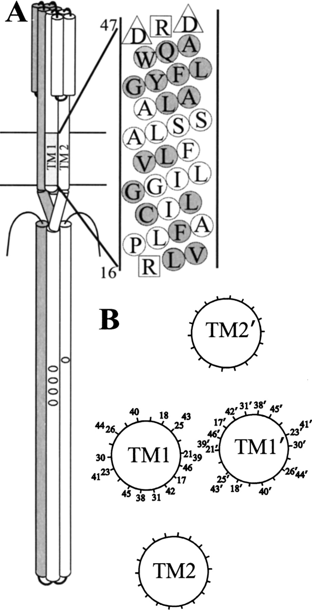
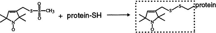
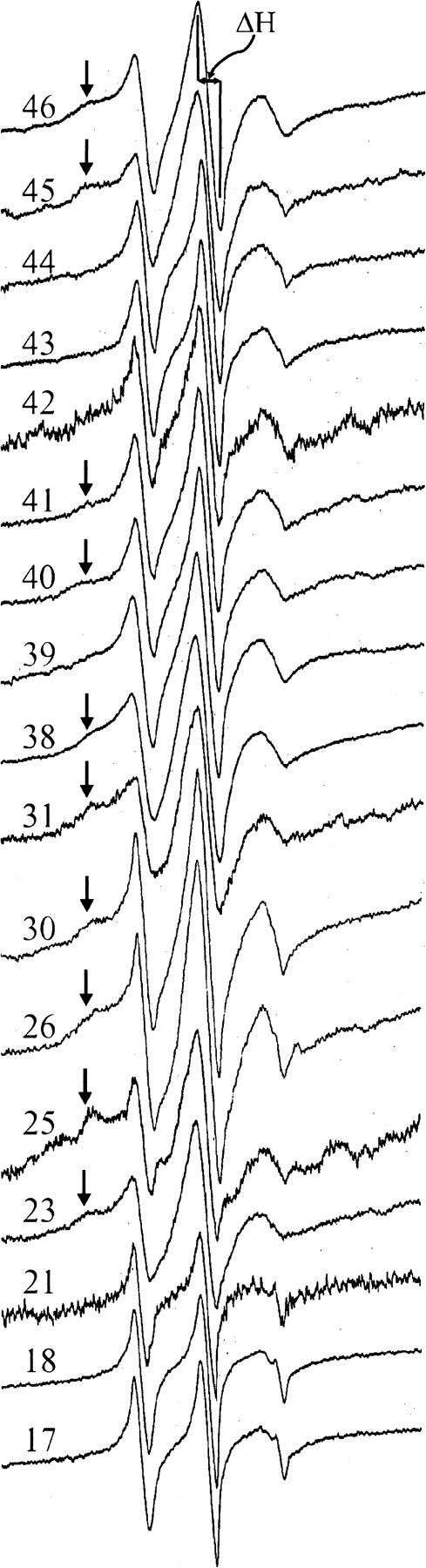
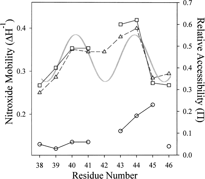
We used site-directed spin labeling and electron paramagnetic resonance spectroscopy to investigate dynamics and helical packing in the four-helix transmembrane domain of the homodimeric bacterial chemoreceptor Trg. We focused on the first transmembrane helix, TM1, particularly on the nine-residue sequence nearest the periplasm, because patterns of disulfide formation between introduced cysteines had identified that segment as the region of closest approach among neighboring transmembrane helices. Along this sequence, mobility and accessibility of the introduced spin label were characteristic of loosely packed or solvent-exposed side chains. This was also the case for eight additional positions around the circumference and along the length of TM1. For the continuous nine-residue sequence near the periplasm, mobility and accessibility varied only modestly as a function of position. We conclude that side chains of TM1 that face the interior of the four-helix domain interact with neighboring helices but dynamic movement results in loose packing. Compared to transmembrane segments of other membrane proteins reconstituted into lipid bilayers and characterized by site-directed spin labeling, TM1 of chemoreceptor Trg is the most dynamic and loosely packed. A dynamic, loosely packed chemoreceptor domain can account for many experimental observations about the transmembrane domains of chemoreceptors.


Figures

Fig. 1.
Fig. 1.
Chemoreceptor organization. (A) Model of a chemoreceptor dimer and details of TM1 of Trg. On the left is a cartoon of a chemoreceptor dimer with one subunit shaded. On one subunit, transmembrane helices 1 (TM1) and 2 (TM2) are labeled and methyl-accepting sites shown as small ovals. On the right, the sequence of TM1 from Trg is shown in a helical net diagram with residue numbers provided for the two bracketing charged residues. Negatively charged residues are indicated by triangles and positively charged residues by squares. Positions characterized by site-directed spin labeling in this study are shaded. (B) Model of the transmembrane domain of Trg showing orientations and relative separations of the four helices determined by cross-linking moments (Lee and Hazelbauer 1995). Positions characterized by site-directed spin labeling in this study are indicated by residue number.
Fig. 2.
Fig. 2.
Reaction of the methanethiosulfonate spin label with a cysteine-containing protein to produce the nitroxide side chain designated R1.
Fig. 3.
Fig. 3.
First-derivative EPR spectra of forms of Trg containing a nitroxide side chain R1 at the indicated positions. The spectra are scaled vertically for convenience of presentation. The magnetic field scan width was 98 G. The line width of the central resonance (ΔH) is indicated on the top spectrum. For spectra that appear to have contributions from two or more components, the less-mobile components are marked by arrows. ΔH−1 values for the spectra shown were: 46, 0.294; 45, 0.284; 44, 0.400; 43, 0.380; 42, 0.345; 41, 0.345; 40, 0.351; 39, 0.286; 38, 0.250; 31, 0.286; 30, 0.394; 26, 0.353; 25, 0.333; 23, 0.312; 21, 0.426; 18, 0.455; 17, 0.455.
Fig. 4.
Fig. 4.
Mobility and accessibility of the R1 spin label along a nine-residue sequence of TM1. The mobility parameter ΔH−1 (▵) and the accessibility parameters |gP(O2) (□) and |gP(NiEDDA) (○) are plotted for positions 38–46 of chemoreceptor Trg. The gray, continuous curve is a plot of the helical periodicity of TM1 derived from cysteine-scanning (Lee et al. 1995a) and patterns of disulfide cross-linking between introduced cysteines (Lee et al. 1994; Hughson et al. 1997), with minima corresponding to the face of TM1 closest to its nearest neighbors TM1` and TM2 and maxima corresponding to the solvent-exposed face (see Fig. 1B ▶).
Fig. 5.
Fig. 5.
Immersion depths for solvent-exposed positions in TM1 of chemoreceptor Trg. Values of depth from the phosphate groups of the lipid bilayer were calculated as described (Gross et al. 1999). The dotted line has a slope of 1.5 Å/residue. The hydrophobic/hydrophilic boundary of the bilayer is at ∼5 Å.
Fig. 6.
Fig. 6.
Mobility and accessibility of interior-facing R1 residues on transmembrane α-helices characterized by site-directed spin labeling. Average values for the mobility parameter Δ H−1 (A) or the accessibility parameter |gP(O2) (B) are shown for interior-facing helical positions of bacteriorhodopsin (bR) (Altenbach et al. 1990, 1994), rhodopsin (Rho) (Altenbach et al. 1996), diphtheria toxin T domain (DT) inserted into bilayers at low pH (Oh et al. 1996), Streptomyces lividans potassium channel (KscA) (Gross et al. 1999), colicin E1 (ColE1) inserted into bilayers at low pH (Salwinski and Hubbell 1999), annexin XII (AXII) inserted into membranes at low pH (Langen et al. 1998), lactose permease (LacY) (Voss et al. 1996), and TM1 of Trg (this study). For each protein, all the sites were along a single helix. The number of values averaged is indicated within the bar and was the same for panels A and B. Error bars represent the standard deviation from the mean. Representative spectra from positions used in the calculations for panel A are shown in panel C.

References

    1. Almeida, F.C. and Opella, S.J. 1997. fd coat protein structure in membrane environments: Structural dynamics of the loop between the hydrophobic trans-membrane helix and the amphipathic in-plane helix. J. Mol. Biol. 270 481–495. - PubMed
    1. Altenbach, C., Cai, K., Khorana, H.G., and Hubbell, W.L. 1999. Structural features and light-dependent changes in the sequence 306–322 extending from helix VII to the palmitoylation sites in rhodopsin: A site-directed spin-labeling study. Biochemistry 38 7931–7937. - PubMed
    1. Altenbach, C., Greenhalgh, D.A., Khorana, H.G., and Hubbell, W.L. 1994. A collision gradient method to determine the immersion depth of nitroxides in lipid bilayers: Application to spin-labeled mutants of bacteriorhodopsin. Proc. Natl. Acad. Sci. 91 1667–1671. - PMC - PubMed
    1. Altenbach, C., Marti, T., Khorana, H.G., and Hubbell, W.L. 1990. Transmembrane protein structure: Spin labeling of bacteriorhodopsin mutants. Science 248 1088–1092. - PubMed
    1. Altenbach, C., Oh, K.J., Trabanino, R.J., Hideg, K., and Hubbell, W.L. 2001. Estimation of inter-residue distances in spin labeled proteins at physiological temperatures: Experimental strategies and practical limitations. Biochemistry 40 15471–15482. - PubMed

Publication types

LinkOut - more resources