. 2002 May 15;16(10):1209-19.
doi: 10.1101/gad.966002.

Oct4 distribution and level in mouse clones: consequences for pluripotency

Affiliations

Oct4 distribution and level in mouse clones: consequences for pluripotency

Michele Boiani et al. Genes Dev. .

Abstract

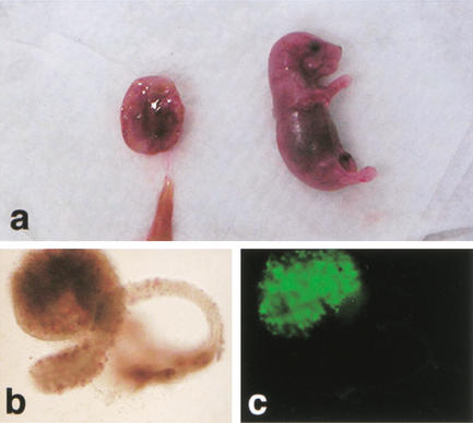
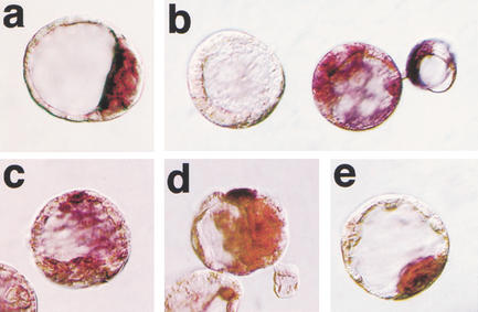
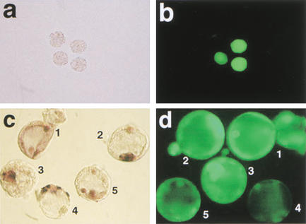
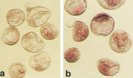
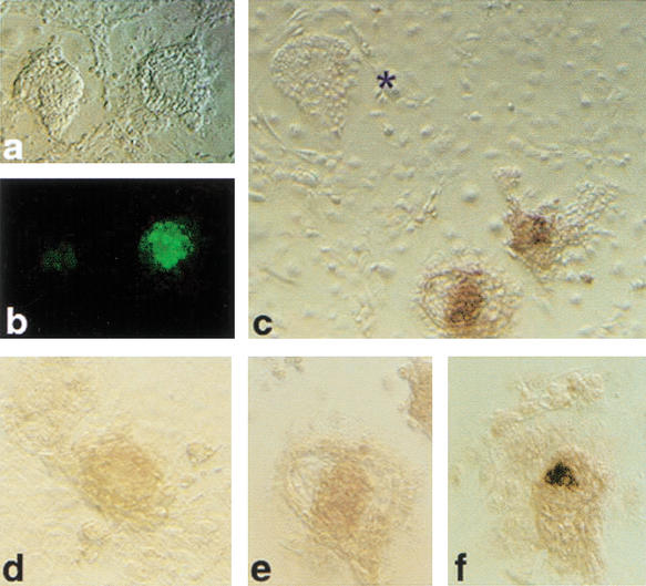
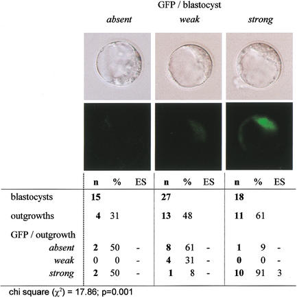
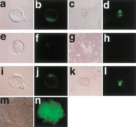
Somatic cell clones often fail at a developmental stage coincident with commencement of differentiation. The transcription factor Oct4 is expressed during cleavage stages and is essential for the differentiation of the blastocyst. Oct4 expression becomes restricted to the inner cell mass and epiblast. After gastrulation Oct4 is active only in germ cells and is silent in somatic cells. Here, Oct4 and an Oct4-GFP transgene were used as markers for which gene reprogramming could be directly related to the developmental potential of somatic cell clones. Cumulus cell clones initiated Oct4 expression at the correct stage but showed an incorrect spatial expression in the majority of blastocysts. The ability of clones to form outgrowths was reduced, and the outgrowths had low or even undetectable levels of Oct4 RNA or GFP. The quality of GFP signals in blastocysts correlated with the ability to generate outgrowths that maintain GFP expression and the frequency of embryonic stem (ES) cell derivation. Abnormal Oct4 expression in clones is either directly or indirectly caused by reprogramming errors and is indicative of a general failure to reset the genetic program. The abnormal Oct4 expression may be associated with aberrant expression of other crucial developmental genes, leading to abnormalities at various embryonic stages. Regardless of other genes, the variations observed in Oct4 levels alone account for the majority of failures currently observed for somatic cell cloning.


Figures

Figure 1
Figure 1
Term development of Oct4–GFP cumulus cell clones. (a) Female pup and placenta recovered 19.5 dpc; (b) Ovary (brightfield); (c) ovary (fluorescence), identifying Oct4–GFP-positive germ cells.
Figure 2
Figure 2
Oct4 mRNA distribution in blastocysts. Whole-mount in situ hybridization. (a) Blastocyst-stage fertilized embryo showing restriction of Oct4 mRNA to the ICM caused by down-regulation of transcription in the TE. (be) Blastocyst-stage cumulus cell clones with lack of expression (b, left embryo), random expression in ICM and trophectoderm (b, right embryo; c, d), and ICM-specific expression (e). The color reaction in samples be was for 3 h to verify lack of signal in negative embryos.
Figure 3
Figure 3
Oct4 and Oct4–GFP in cumulus cell clones at preimplantation stages. (a,b) Oct4–GFP expression in cumulus cell clones at the morula stage. (a) Bright field morphology, (b) GFP fluorescence with examples of clones both expressing and lacking Oct4–GFP. (c,d) Whole-mount in situ hybridization for Oct4 mRNA (c) and GFP fluorescence (d) in clones at the blastocyst stage. Consistency between Oct4–GFP and Oct4 in situ signal is observed in embryos 2 and 3, whereas embryo 4 lacks GFP signal but shows strong Oct4 expression in trophectoderm cells. Embryos 1 and 5 lack an ICM-specific signal.
Figure 4
Figure 4
Expression level of Oct4 in cumulus cell clones and fertilized blastocysts detected by in situ hybridization. Processing of embryos in the reaction was simultaneous, documentation after 1 h of color reaction. (a) Blastocyst-stage cumulus cell clones; (b) fertilized controls.
Figure 5
Figure 5
Outgrowths from cumulus cell clones after 3 d of culture on a feeder layer. (a) Bright field; (b) Oct4–GFP fluorescence on same outgrowths as in a; (c) in situ hybridization for Oct4 mRNA on synchronous clone outgrowths; (*) outgrowth lacking Oct4 mRNA-expressing cells. (d,e) Clone outgrowths and (f) outgrowth from fertilized blastocyst illustrating level of Oct4 expression detected after 2 h of color reaction.
Figure 6
Figure 6
Oct4–GFP level in clone blastocysts and outgrowth formation.
Figure 7
Figure 7
Oct4–GFP in blastocyst-stage clones, outgrowths, and ES cells. (ad) Fertilized embryo and (el) clones at the blastocyst stage and their subsequent outgrowth; (m,n) ES cell formation from the last embryo illustrated in il including corresponding GFP signals. The clone in e and f is almost negative for GFP, and shows no GFP in the outgrowths. The clone in i and j has GFP comparable to the control and shows GFP-positive cells in both ICM and outgrowth, and formed ES cells.

Comment in

  • Cloning v. clowning.
    Solter D. Solter D. Genes Dev. 2002 May 15;16(10):1163-6. doi: 10.1101/gad.998302. Genes Dev. 2002. PMID: 12023296 No abstract available.

References

    1. Abbondanzo SJ, Gadi I, Stewart CL. Derivation of embryonic stem cell lines. Methods Enzymol. 1993;225:803–823. - PubMed
    1. Amano T, Tani T, Kato Y, Tsunoda Y. Mouse cloned from embryonic stem (ES) cells synchronized in metaphase with nocodazole. J Exp Zool. 2001;289:139–145. - PubMed
    1. Botquin V, Hess H, Fuhrmann G, Anastassiadis C, Gross MK, Vriend G, Schöler HR. New POU dimer configuration mediates antagonistic control of an osteopontin preimplantation enhancer by Oct-4 and Sox-2. Genes & Dev. 1998;12:2073–2090. - PMC - PubMed
    1. Chawengsaksophak K, James R, Hammond VE, Kontgen F, Beck F. Homeosis and intestinal tumours in Cdx2 mutant mice. Nature. 1997;386:84–87. - PubMed
    1. Cheong HT, Takahashi Y, Kanagawa H. Birth of mice after transplantation of early cell-cycle-stage embryonic nuclei into enucleated oocytes. Biol Reprod. 1993;48:958–963. - PubMed

MeSH terms

LinkOut - more resources