. 2002 May 15;16(10):1234-46.
doi: 10.1101/gad.222902.

Cooperative action of Tbx2 and Nkx2.5 inhibits ANF expression in the atrioventricular canal: implications for cardiac chamber formation

Affiliations

Cooperative action of Tbx2 and Nkx2.5 inhibits ANF expression in the atrioventricular canal: implications for cardiac chamber formation

Petra E M H Habets et al. Genes Dev. .

Abstract

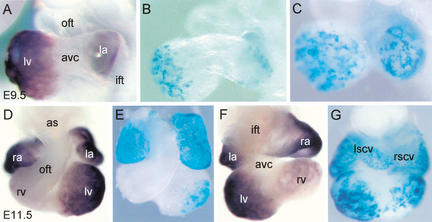
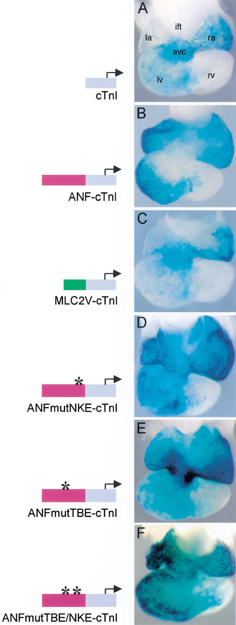
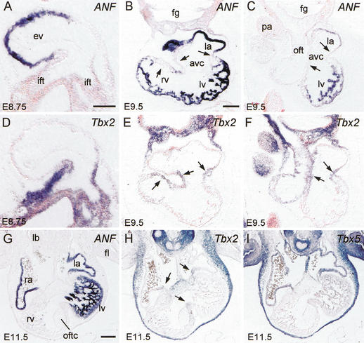
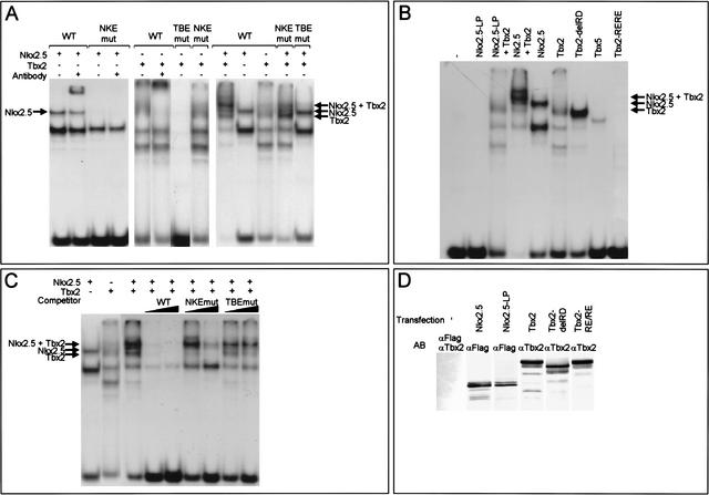
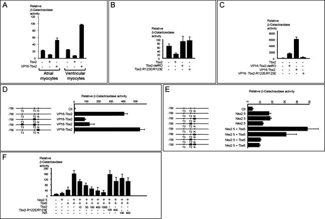
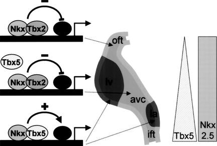
During heart development, chamber myocardium forms locally from the embryonic myocardium of the tubular heart. The atrial natriuretic factor (ANF) gene is specifically expressed in this developing chamber myocardium and is one of the first hallmarks of chamber formation. We investigated the regulatory mechanism underlying this selective expression. Transgenic analysis shows that a small fragment of the ANF gene is responsible for the developmental pattern of endogenous ANF gene expression. Furthermore, this fragment is able to repress cardiac troponin I (cTnI) promoter activity selectively in the embryonic myocardium of the atrioventricular canal (AVC). In vivo inactivation of a T-box factor (TBE)- or NK2-homeobox factor binding element (NKE) within the ANF fragment removed the repression in the AVC without affecting its chamber activity. The T-box family member Tbx2, encoding a transcriptional repressor, is expressed in the embryonic myocardium in a pattern mutually exclusive to ANF, thus suggesting a role in the suppression of ANF. Tbx2 formed a complex with Nkx2.5 on the ANF TBE-NKE, and was able to repress ANF promoter activity. Our data provide a potential mechanism for chamber-restricted gene activity in which the cooperative action of Tbx2 and Nkx2.5 inhibits expression in the AVC.


Figures

Figure 1
Figure 1
The 0.7-kb ANF regulatory region is responsible for the developmental pattern of the endogenous ANF gene. (A) A lateral view of the endogenous ANF gene expression in the heart of E9.5 mouse embryo. (B,C) A lateral view of the nlacZ reporter gene expression in the heart of E9.5 transgenic embryos. (D,F) A ventral (D) and dorsal (F) view of the endogenous ANF expression at E11.5. (E,G) A ventral (E) and dorsal (G) view of ANF transgene expression at E11.5. (ift) Inflow tract; (la) left atrium; (ra) right atrium; (avc) atrioventricular canal; (lv) left ventricle; (rv) right ventricle; (oft) outflow tract; (lscv) left superior caval vein; (rscv) right superior caval vein; (as) aortic sac.
Figure 2
Figure 2
. Localization of transgene expression in E10.5 mouse hearts. (A) The cTnI transgene is predominantly expressed in the primary myocardium of the AVC. (B) The ANF–cTnI transgene is solely expressed in the chamber myocardium. No transgene expression is present in the AVC myocardium. (C) MLC2V–cTnI transgenics show predominant expression in the primary myocardium of the AVC, similar to the pattern of the cTnI transgenes. (D) Mutation of the NKE at position −250 bp in the ANF regulatory region removes the repression in the AVC. (E) Mutation of the TBE at position −259 bp in the ANF regulatory region also removes the repression in the AVC. (F) Mutation of both the TBE and NKE removes the repression in the AVC as well. (ift) Inflow tract; (la) left atrium; (ra) right atrium; (avc) atrioventricular canal; (lv) left ventricle; (rv) right ventricle; (oft) outflow tract.
Figure 3
Figure 3
Nonradioactive in situ hybridization on serial sections shows complementary expression of endogenous ANF, Tbx2, and Tbx5 mRNA. (A–C,G) ANF expression at E8.75 (A), E9.5 (B,C), and E11.5 (G). (D–F,H) Tbx2 expression at E8.75 (D), E9.5 (E,F), and E11.5 (H). (I) Tbx5 expression at E11.5. Note the mutually exclusive pattern of expression of ANF and Tbx2. Arrows in B and E indicate the AVC and OFT region that is continuous at the inner curvature. Arrows in C, F, and H indicate the AVC region. (ift) Inflow tract; (la) left atrium; (ra) right atrium; (avc) atrioventricular canal; (lv) left ventricle; (rv) right ventricle; (oft) outflow tract; (ev) embryonic ventricle; (fg) foregut; (pa) pharyngeal arch; (oftc) outflow tract cushion; (lb) lung bud; (fl) forelimb. Bar, 100 μm.
Figure 4
Figure 4
Tbx2 and Nkx2.5 form a ternary complex with the ANF TBE–NKE. (A) EMSAs were performed using nuclear extracts from HEK cells expressing Nkx2.5 or Tbx2. The wild-type probe contains the TBE–NKE, the NKEmut probe contains the TBE, and the mutated NKE as used in the transgene construct. The TBEmut probe contains the NKE and the mutated TBE as used in the transgene construct. Both Nkx2.5 and Tbx2 bind to the wild-type probe. Nkx2.5 does not bind the NKEmut probe and Tbx2 does not bind the TBEmut probe. When mixing together Nkx2.5 and Tbx2 extracts, an additional ternary complex is formed on the wild type, and to a lesser extent, on the NKEmut probe, whereas the complex is absent when using the TBEmut probe. (B) Replacing Nkx2.5 for Nkx2.5-L176P (NKx2.5-LP) or Tbx2 for Tbx2-R122E/R123E (Tbx2-RE/RE) shows that on the wild-type probe, the DNA-binding ability of both Nkx2.5 and Tbx2 is necessary for complex formation. The carboxy-terminal region of Tbx2 is not required for DNA binding (Tbx2-delRD). Tbx5 is also able to bind the wild-type probe. (C) Once formed, the Nkx2.5/Tbx2 complex is stable. The wild-type probe was incubated with nuclear extracts and cold competitor oligonucleotides (100- and 1000-fold excess) as indicated at top. Whereas a 100-fold excess of wild-type probe was sufficient to disrupt the complex, a 100-fold excess of NKEmut and a 1000-fold excess of TBEmut were not sufficient. (D) Western blots of nuclear extracts of HEK cells expressing FLAG–Nkx2.5, FLAG–Nkx2.5-LP, Tbx2, Tbx2-delRD and Tbx2-RE/RE.
Figure 5
Figure 5
The 0.7-kb ANF promoter is a functional target for Tbx2. (A) Transient transfections were carried out with the 0.7-kb ANF promoter in primary atrial and ventricular cardiomyocytes. Cotransfections show that Tbx2 repressed ANF promoter activity, whereas VP16-Tbx2-delRD activated the ANF promoter. The results are from one representative experiment (out of 3) done in duplicate. Error bars represent the difference between the duplicates. (B) Tbx2-mediated repression requires the Tbx2 repressor domain and interaction with the DNA. Cotransfection experiments were carried out with the 0.7-kb ANF promoter in Cos-7 cells. (C) Fusion of the VP16 transactivation domain to either the full-length Tbx2 (VP16-Tbx2) or to Tbx2, from which the carboxy-terminal end that includes the repression domain, was removed (VP16-Tbx2-delRD) resulted in strong activation of the ANF promoter region. VP16-Tbx2-R122E/R123E did not activate the ANF promoter region. (D) Cotransfection, using point mutations of the 0.7-kb ANF promoter in Cos-7 cells, shows that VP16-Tbx2 activates the ANF regulatory region via the TBE. The basal values of the mutated constructs were comparable with the control construct. (E) Nkx2.5 activation of the ANF promoter does not require the NKE. Synergistic activation of the ANF promoter by Tbx5 and Nkx2.5 requires an intact TBE and NKE. The basal values of the mutated constructs were comparable with the control construct. (F) Synergistic activity of Nkx2.5 and Tbx5 is reduced by Tbx2 in a dose-dependent manner, indicating that Tbx2 can efficiently compete with Tbx5 in the regulation of the ANF promoter. All results are from one representative experiment (out of 3) done in duplicate. Error bars represent the difference between the duplicates. (N) NKE located at position −250 bp; (T2) TBE located at position −259 bp; (T3) TBE located at position −485 bp.
Figure 6
Figure 6
A potential mechanism for site-specific chamber formation by local repression of differentiation. Schematic representation of the transcriptional mechanisms involved in chamber formation. As part of an ongoing chamber formation program, Tbx5 and Nkx2.5 stimulate cardiac genes. Specific regions in the linear heart tube remain embryonic and do not develop into chamber myocardium due to the presence of Tbx2 in those regions. Nkx2.5 and Tbx2 form a repressor complex that suppresses genes that are part of the chamber differentiation program. The Tbx5 triangle and Nkx2.5 rectangle indicate Tbx5 and Nkx2.5 expression in the linear heart tube, respectively. Tbx2 is expressed in the primary myocardium of the inflow tract, atrioventricular canal, and outflow tract (light gray), whereas ANF is expressed in the chamber myocardium (dark gray). (ift) Inflow tract; (la) left atrium; (avc) atrioventricular canal; (lv) left ventricle; (oft) outflow tract.

References

    1. Argentin S, Ardati A, Tremblay S, Lihrmann I, Robitaille L, Drouin J, Nemer M. Developmental stage-specific regulation of atrial natriuretic factor gene transcription in cardiac cells. Mol Cell Biol. 1994;14:777–790. - PMC - PubMed
    1. Ausoni S, de Nardi C, Moretti P, Gorza L, Schiaffino S. Developmental expression of rat cardiac troponin I mRNA. Development. 1991;112:1041–1051. - PubMed
    1. Basson CT, Bachinsky DR, Lin RC, Levi T, Elkins JA, Soults J, Grayzel D, Kroumpouzou E, Traill TA, Leblanc-Straceski J, et al. Mutations in human TBX5 (corrected) cause limb and cardiac malformation in Holt-Oram syndrome. Nat Genet. 1997;15:30–35. - PubMed
    1. Basson CT, Huang T, Lin RC, Bachinsky DR, Weremowicz S, Vaglio A, Bruzzone R, Quadrelli R, Lerone M, Romeo G, et al. Different TBX5 interactions in heart and limb defined by Holt-Oram syndrome mutations. Proc Natl Acad Sci. 1999;96:2919–2924. - PMC - PubMed
    1. Bell AC, West AG, Felsenfeld G. The protein CTCF is required for the enhancer blocking activity of vertebrate insulators. Cell. 1999;98:387–396. - PubMed

Publication types

MeSH terms