. 2002 May 15;16(10):1247-59.
doi: 10.1101/gad.969702.

Two interacting bZIP proteins are direct targets of COP1-mediated control of light-dependent gene expression in Arabidopsis

Affiliations

Two interacting bZIP proteins are direct targets of COP1-mediated control of light-dependent gene expression in Arabidopsis

Magnus Holm et al. Genes Dev. .

Abstract

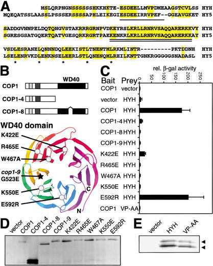
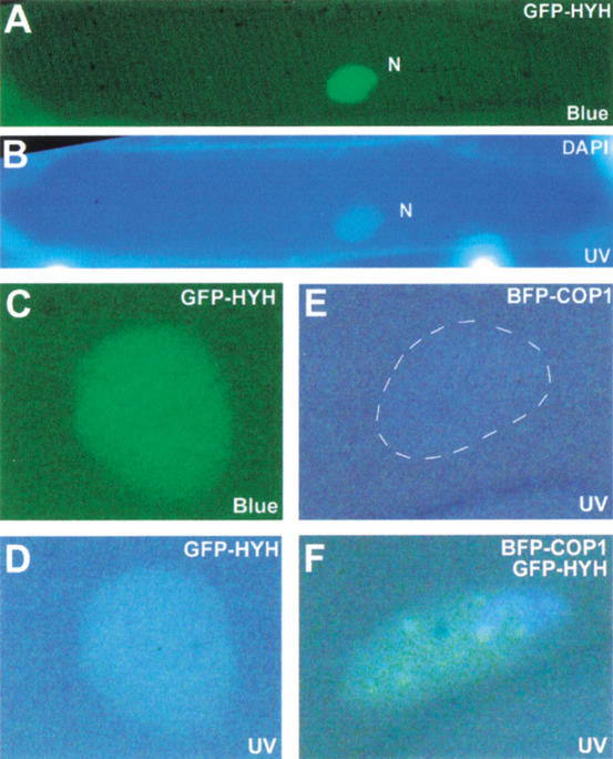
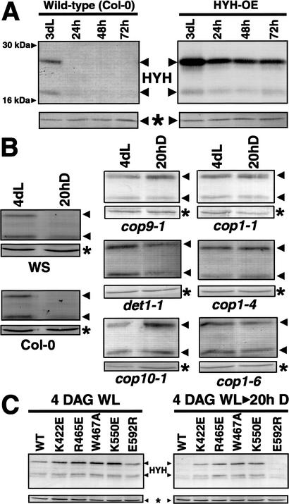
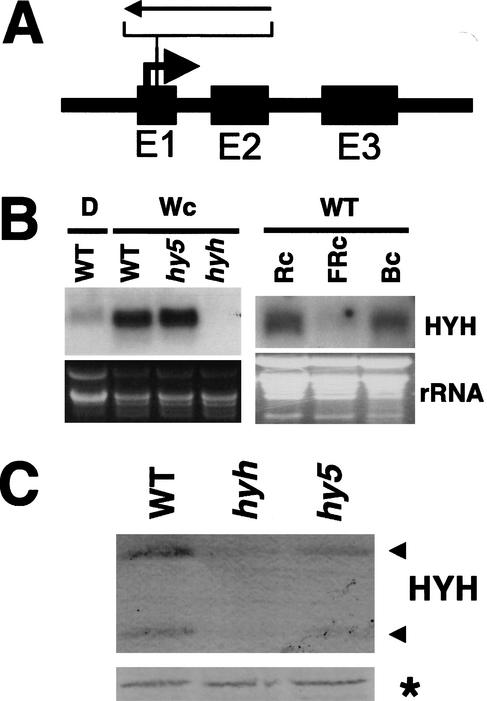
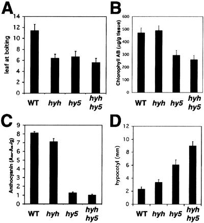
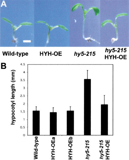
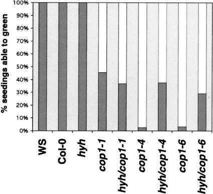
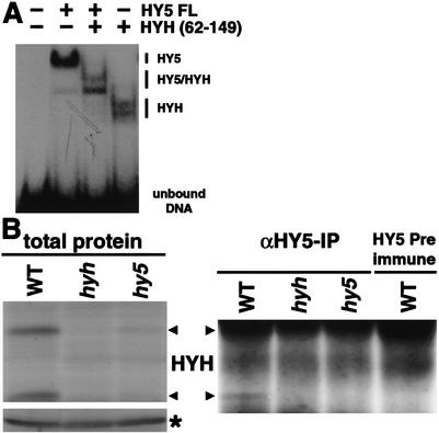
Arabidopsis COP1 acts to repress photomorphogenesis in the absence of light. It was shown that in the dark, COP1 directly interacts with the bZIP transcription factor HY5, a positive regulator of photomorphogenesis, and promotes its proteasome-mediated degradation. Here we identify a novel bZIP protein HYH, as a new target of COP1. We identify a physical and genetic interaction between HYH and COP1 and show that this interaction results in dark-specific degradation of HYH. Genetic analysis indicates that HYH is predominantly involved in blue-light regulation of development and gene expression, and that the function of HYH in part overlaps with that of HY5. The accumulation of HYH protein, not the mRNA, is dependent on the presence of HY5. Our data suggest that HYH and HY5 can, respectively, act as heterodimers and homodimers, thus mediating light-regulated expression of overlapping as well as distinct target genes. We propose that COP1 mediates light control of gene expression through targeted degradation of multiple photomorphogenesis-promoting transcription factors in the nucleus.


Figures

Figure 1
Figure 1
HYH interacts with the WD40 domain of COP1 through a COP1-interaction motif. (A) Alignment of HYH and HY5; identical amino acids are shaded in yellow. The COP1-interaction motifs (Holm et al. 2001) are single underlined and the conserved DNA-binding basic domains are double underlined; leucines in the dimerizing leucine zipper are marked by asterisks. (B) Domain organization and mutations in COP1. The genetically identified mutations cop1-4, cop1-8, and cop1-9, as well as the amino-acid-substituted COP1 proteins have been previously described (Holm et al. 2001). The ribbon diagram of the Gβ β-propeller was used to align the COP1 WD40 repeats. The carbon α backbone (Cα) positions of the substituted COP1 residues are marked by circles. The star marks the position of the cop1-9 mutation. (C) Yeast two-hybrid interactions between wild-type or mutated COP1 proteins and HYH proteins. VP-AA indicates alanine substitution of the conserved VP pair in the COP1-interacting motif of HYH. Error bar represent standard deviation, n = 6. (D,E) Immunoblots of yeast expressing indicated Gal4 DNA-binding domain fused COP1 and activation-domain fused HYH proteins, respectively.
Figure 2
Figure 2
HYH is nuclear and colocalizes with COP1 in onion cells. (A) Blue-light excitation of a DAPI-stained onion epidermal cell expressing GFP–HYH. The diffuse GFP fluorescence is restricted to the nucleus (N). (B) The same cell as in A excited by UV light to show DAPI stain to indicate position of the nucleus. (C) A nucleus expressing GFP–HYH excited by blue light. (D) The nucleus in C excited by UV light. (E) A nucleus expressing BFP–COP1 excited by UV light. (F) A representative onion cell nucleus that expresses both GFP–HYH and BFP–COP1 showing FRET-induced green speckles upon UV light excitation. FRET can occur when BFP and GFP proteins are in close proximity; blue photons emitted by UV-excited BFP can then excite GFP to allow emission of green light.
Figure 3
Figure 3
The COP1 interaction results in degradation of HYH in the dark. (A) Both endogenous and overexpressed HYH protein is degraded upon transfer to darkness. The wild-type (Col-0) and HYH-OE seedlings were grown in white light for 3 d and then transferred to darkness. Protein extracts were prepared at day 3 and after 24, 48, and 72 h in darkness, and the protein concentration was normalized before loading. Neither of the 27- and 21-kD bands were detected using preimmune sera (data not shown). The HYH proteins migrate distinct from the 30-kD HY5 protein, and no cross-reactivity to endogenous HY5 was observed in immunoblots. (B) Wild-type (Col-0 and WS ecotypes) and cop9-1, det1-1, cop10-1, cop1-1, cop1-4, and cop1-6 seedlings were grown in white light for 4 d and for an additional 20 h in darkness, respectively. Protein extracts were prepared at 4 DAG and at 4 DAG + 20 h, and the protein concentration was normalized before loading. (C) Seedlings overexpressing wild-type or indicated amino-acid-substituted COP1 protein in a cop1-5 null mutant background were grown in white light for 4 d and then transferred to darkness for 20 h. Protein extracts were prepared at day 4 (4 DAG), and after 20 h in darkness (20h D), the protein concentration was normalized and 15 μg of total extract was loaded in each lane. The amount of HYH protein was assayed by immunoblotting; the asterisk marks a cross-reacting band that serves as an internal loading control. At least two independent lines were used for each transgenic construct, and similar results were observed.
Figure 4
Figure 4
Identification of a knockout mutation in the HYH gene. (A) Schematic illustration of the HYH gene organization with the position of the T-DNA insertion indicated. (B) Northern blot of 20 μg of total RNA prepared from 6-DAG dark-grown wild-type (WS) seedlings (D), and 6-DAG wild-type (WS), hy5-ks50, and hyh seedlings grown in white light, respectively. Right panel shows Northern blot of 20 μg of total RNA prepared from 6-DAG wild-type (Col-0) seedlings grown in continuous red (108 μmole/sec per m2), far-red (160 μmole/sec per m2), and blue (16 μmole/sec per m2) light, respectively. The HYH RNA is detected as a single band at ∼600 bp. (Bottom panels) The ethidium bromide-stained gel serving as a loading control. (C) Immunoblot of 10 μg of total protein prepared from 3-DAG wild-type (WS), hyh, and hy5-ks50 seedlings grown in white light. The two proteins detected with the HYH antibodies are indicated with arrows to the right. The asterisk marks a cross-reacting protein serving as a loading control.
Figure 5
Figure 5
Characterization of the hyh mutant. (A) Number of rosette leaves at bolting for wild-type, hyh, hy5-ks50, and hyh/hy5-ks50 plants grown in long-day conditions (16 h light, 8 h dark). (B) Chlorophyll a and b content in 4-DAG wild-type, hyh, hy5-ks50, and hyh/hy5-ks50 seedlings grown in white light. Error bars represent standard deviation, n = 5. (C) Anthocyanin content in 3-DAG wild-type, hyh, hy5-ks50, and hyh/hy5-ks50 seedlings grown in white light. Error bars represent standard deviation, n = 4. (D) Hypocotyl length of 6-DAG wild-type, hyh, hy5-ks50, and hyh/hy5-ks50 seedlings grown in blue light (∼16 μmole/sec per m2). Error bar represents standard deviation, n = 20.
Figure 6
Figure 6
Overexpression of HYH is able to suppress the phenotype of hy5-215. (A) 6-DAG white-light-grown wild-type, HYH-OE, hy5-215, and hy5-215/HYH-OE seedlings. Bar, 1 mm. (B) Graphical representation of the hypocotyl length of the seedlings in A. The columns represent wild-type (WT), two independent transgenic lines overexpressing HYH (HYH-OEa and b), hy5-215 seedlings, and hy5-215 seedlings overexpressing HYH, all in the Col-0 ecotype. Error bars represent standard deviation, n = >30.
Figure 7
Figure 7
The hyh mutation suppresses the block-of-greening phenotype of cop1 alleles. Seedlings were germinated in the dark for 4 d and then transferred to constant white light for 4 d. Seedlings with green cotyledons and/or true leaves were scored as able to green, and bleached seedlings were scored as unable to green (n = >109).
Figure 8
Figure 8
HYH interacts with HY5 on DNA and in vivo. (A) Gel retardation using recombinant HY5 and HYH protein binding to the G-box of the RBCS promoter. Approximately 40 ng each of the HY5 and HYH protein were mixed and incubated at 50°C for 5 min to dissociate pre-existing dimers prior to addition to radioactively labeled probe. The protein–DNA complexes were resolved on a 4% native 0.5× TBE polyacrylamide gel. (B) HY5 immunoprecipitation of 3-DAG wild-type, hyh, and hy5-ks50 seedlings grown in white light. (Left panel) Total protein extract, (*) a cross-reacting protein band serving as loading control, and (right panel) the anti-HY5 precipitate following immunodetection with HYH antibodies. The right lane shows the HY5 preimmune precipitate from wild-type extract.
Figure 9
Figure 9
A functional overlap between HYH and HY5. (A) Hierarchical clustering display of the expression ratios from 6-DAG wild type (WT) seedlings versus hy5-ks50, hyh, or hyh/hy5-ks50 (dbl) seedlings, all grown in blue light. The 149 genes (http://www.plantgenomics.biology.yale.edu/) that showed at least twofold differential change in at least one of the three sample pairs were included for comparison. (B) The differential expression of nine different genes in the hy5, hyh, and hy5/hyh experiments. See text for predicted function and domains of the nine displayed genes. The ratio of wild-type/mutant is expressed as log2(ratio). (C) Venn diagrams of the number of differentially expressed genes that showed twofold or higher expression in wild-type (repressed-red) or lower (induced-green) compared with respective mutant. The numbers in the overlapping areas indicate the shared number of genes showing twofold or higher differential expression in either two or three sample pairs.

References

    1. Ang LH, Deng XW. Regulatory hierarchy of photomorphogenic loci: Allele-specific and light-dependent interaction between the HY5 and COP1 loci. Plant Cell. 1994;6:613–628. - PMC - PubMed
    1. Ang LH, Chattopadhyay S, Wei N, Oyama T, Okada K, Batschauer A, Deng XW. Molecular interaction between COP1 and HY5 defines a regulatory switch for light control of Arabidopsis development. Mol Cell. 1998;1:213–222. - PubMed
    1. The Arabidopsis Genome Initiative. Analysis of the genome sequence of the flowering plant Arabidopsis thaliana. Nature. 2000;408:796–815. - PubMed
    1. Ballesteros ML, Bolle C, Lois LM, Moore JM, Vielle-Calzada JP, Grossniklaus U, Chua NH. LAF1, a MYB transcription activator for phytochrome A signaling. Genes & Dev. 2001;15:2613–2625. - PMC - PubMed
    1. Chattopadhyay S, Ang LH, Puente P, Deng XW, Wei N. Arabidopsis bZIP protein HY5 directly interacts with light-responsive promoters in mediating light control of gene expression. Plant Cell. 1998;10:673–683. - PMC - PubMed

Publication types

MeSH terms