. 2002 Jun;22(12):4124-35.
doi: 10.1128/MCB.22.12.4124-4135.2002.

Type 1 phosphatase, a negative regulator of cardiac function

Affiliations

Type 1 phosphatase, a negative regulator of cardiac function

Andrew N Carr et al. Mol Cell Biol. 2002 Jun.

Abstract

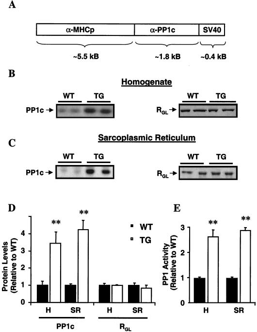
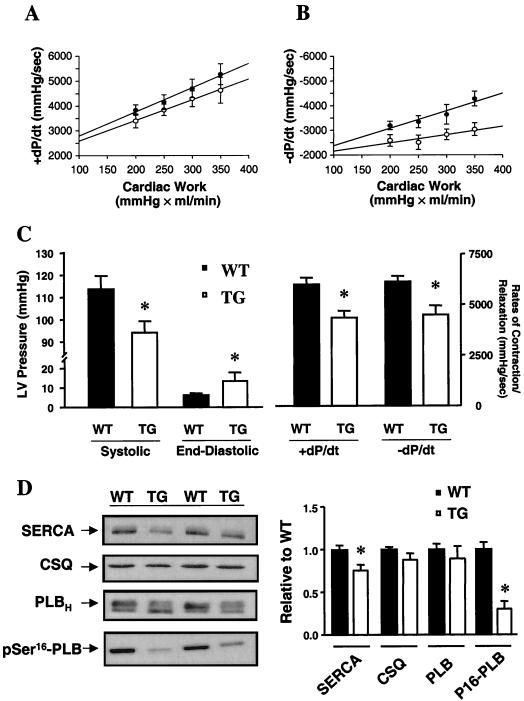
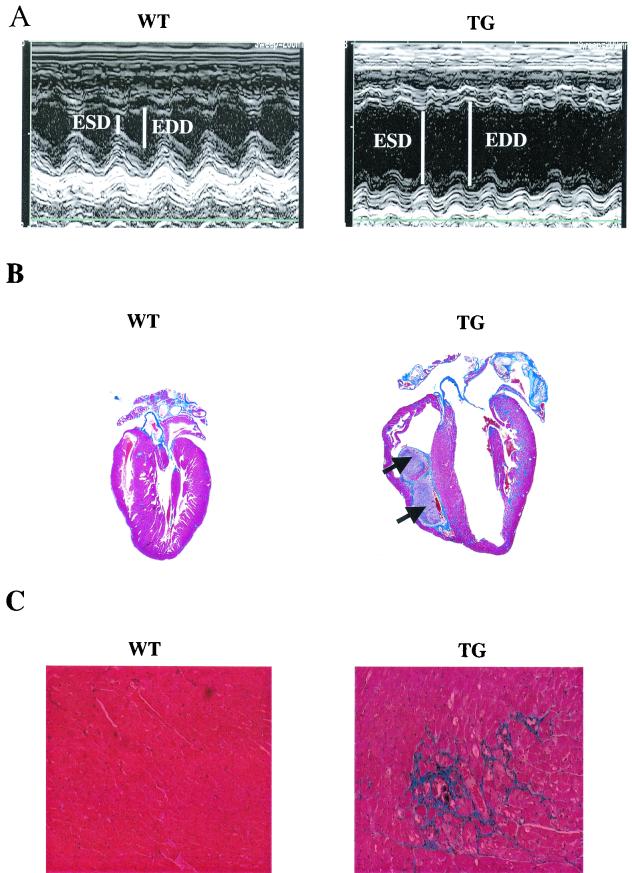
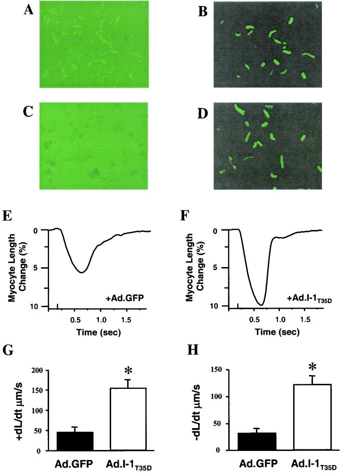
Increases in type 1 phosphatase (PP1) activity have been observed in end stage human heart failure, but the role of this enzyme in cardiac function is unknown. To elucidate the functional significance of increased PP1 activity, we generated models with (i) overexpression of the catalytic subunit of PP1 in murine hearts and (ii) ablation of the PP1-specific inhibitor. Overexpression of PP1 (threefold) was associated with depressed cardiac function, dilated cardiomyopathy, and premature mortality, consistent with heart failure. Ablation of the inhibitor was associated with moderate increases in PP1 activity (23%) and impaired beta-adrenergic contractile responses. Extension of these findings to human heart failure indicated that the increased PP1 activity may be partially due to dephosphorylation or inactivation of its inhibitor. Indeed, expression of a constitutively active inhibitor was associated with rescue of beta-adrenergic responsiveness in failing human myocytes. Thus, PP1 is an important regulator of cardiac function, and inhibition of its activity may represent a novel therapeutic target in heart failure.


Figures

FIG. 1.
FIG. 1.
Generation of PP1 overexpression mice and their characterization at 3 months of age. (A) Cardiac tissue-specific overexpression of the α-isoform of the catalytic subunit of PP1 was driven by the α-MHC promoter. SV40, simian virus 40. (B and C) Representative Western blots of cardiac homogenates (B) and SR preparations (C) revealed similar increases in PP1c protein levels (left) in TG hearts without alterations in RGL, the SR targeting subunit (right). (D) Quantitation of PP1c and RGL levels (left) in whole-heart homogenates (H; n = 5) and SR preparations (SR; n = 6) in TG relative to WT hearts. (E) Quantitation of PP1 activity in homogenates and SR preparations from TG hearts relative to activity in those from WT hearts. Phosphatase activity was 1.3 ± 0.1 nmol/min/mg (n = 5) for the WT homogenates and 3.6 ± 0.3 nmol/min/mg (n = 5) for the TG homogenates and 0.21 ± 0.01 nmol/min/mg (n = 6) for WT SR preparations and 0.61 ± 0.02 nmol/min/mg (n = 6) for TG SR preparations. ∗∗, P < 0.001 versus WT.
FIG. 2.
FIG. 2.
Cardiac function in PP1 mice at 3 months of age. (A and B) Bivariate regression plots of the positive (A; +dP/dt) and negative (B; −dP/dt) first derivatives of intraventricular pressure versus cardiac work in five WT (•) and five TG (○) isolated work-performing hearts. (C) Systolic pressure, end-diastolic pressure, and +dP/dt and −dP/dt in isoproterenol-stimulated perfused hearts. (D) Western blots of SERCA (6 μg), calsequestrin (CSQ; 6 μg), phospholamban (PLB; 6 μg), and pSer16-PLB (10 μg) in individual WT and TG hearts. Immunoreactivity for each protein was compared to a linear standard (2, 4, 8, and 12 μg) consisting of five pooled WT hearts on each blot. Quantitation revealed decreases in SERCA and pSer16-PLB in TG hearts (n = 5) compared to WT hearts (n = 5). ∗, P < 0.05 versus WT.
FIG. 3.
FIG. 3.
PP1 is associated with pathology at 6 months of age. (A) In vivo, M-mode echocardiography performed in hearts from 6-month-old TG mice (right) revealed increased end-systolic (ESD) and end-diastolic dimensions (EDD) indicating LV dilation, compared to results for age-matched WT mice (left). (B) Masson's trichrome-stained longitudinal sections from WT (left) and TG (right) hearts showed dilated cardiomyopathy and the presence of intracardiac thrombi (arrows) in TG hearts. (C) Higher magnification (×170) indicated widespread interstitial fibrosis (blue) in the TG, but not WT, hearts. (D) Representative dot blot of ventricular gene expression showed increases in atrial natriuretic factor (ANF), β-MHC, and α-skeletal actin (α-sk. actin) in TG hearts. Blots are from three animals in each group. GAPDH, glyceraldehyde-3-phosphate dehydrogenase. (E) Survival curves indicated premature mortality (P < 0.00001) in TG (n = 46) compared to WT (n > 100) mice. Survival statistics were obtained by using the log rank test. Cum., cumulative.
FIG. 3.
FIG. 3.
PP1 is associated with pathology at 6 months of age. (A) In vivo, M-mode echocardiography performed in hearts from 6-month-old TG mice (right) revealed increased end-systolic (ESD) and end-diastolic dimensions (EDD) indicating LV dilation, compared to results for age-matched WT mice (left). (B) Masson's trichrome-stained longitudinal sections from WT (left) and TG (right) hearts showed dilated cardiomyopathy and the presence of intracardiac thrombi (arrows) in TG hearts. (C) Higher magnification (×170) indicated widespread interstitial fibrosis (blue) in the TG, but not WT, hearts. (D) Representative dot blot of ventricular gene expression showed increases in atrial natriuretic factor (ANF), β-MHC, and α-skeletal actin (α-sk. actin) in TG hearts. Blots are from three animals in each group. GAPDH, glyceraldehyde-3-phosphate dehydrogenase. (E) Survival curves indicated premature mortality (P < 0.00001) in TG (n = 46) compared to WT (n > 100) mice. Survival statistics were obtained by using the log rank test. Cum., cumulative.
FIG. 4.
FIG. 4.
Protein phosphatase I-1 in the heart. (A and B) Rates of cardiac contraction (A) and relaxation (B) obtained in isoproterenol-stimulated work-performing hearts from WT (n = 5) and knockout (KO; n = 7) mice. (C and D) PP1 activity and cAMP levels in perfused hearts. (E) Representative immunoblots (left) and quantitation (right) of SERCA (6 μg), calsequestrin (CSQ; 6 μg), and phospholamban (PLB; 6 μg) protein levels and PKA phosphorylation of PLB (pSer16-PLB; 10 μg) in perfused hearts. Immunoreactivity for each protein was compared to a linear standard (2, 4, 8, and 12 μg) consisting of five pooled WT hearts on each blot. ∗, P < 0.05 versus WT.
FIG. 5.
FIG. 5.
I-1 in human heart failure. (A) Representative immunoblots of the protein levels (top) and phosphorylation (middle) of I-1 in 9 donor (D) and 10 failing (F) heart homogenates. The calsequestrin levels (CSQ; bottom) in the same blots were assessed as an internal control. (B) Quantitation of I-1 protein levels revealed no alterations. However, I-1 phosphorylation in failing hearts was significantly decreased. ∗, P < 0.05 versus donor hearts.
FIG. 6.
FIG. 6.
Adenovirus expression of a constitutively active I-1 protein in cardiomyocytes from failing human hearts. (A to D) Isolated failing human myocytes expressing either β-galactosidase-GFP (Ad.GFP; top) or I-1T35D-GFP (Ad.I-1T35D) were visualized with direct light (left) or fluorescent light (right). Successfully infected cells appear green. (E and F) Representative traces of cardiomyocyte cell shortening in Ad.GFP- (E) and Ad.I-1T35D-infected (F) cells in response to a maximal concentration of isoproterenol (100 nM). (G and H) Rates of cell shortening (G) and relengthening (H) in Ad.GFP- and Ad.I-1T35D-infected cells in response to isoproterenol. Values are averages of at least 8 to 12 cells from three to five human hearts. ∗, P < 0.05.

References

    1. Ahmad, Z., F. J. Green, H. S. Subuhi, and A. M. Watanabe. 1989. Autonomic regulation of type 1 protein phosphatase in cardiac muscle. J. Biol. Chem. 264:3859-3863. - PubMed
    1. Allen, P. B., O. Hvalby, V. Jensen, M. L. Errington, M. Ramsay, F. A. Chaudhry, T. V. Bliss, J. Storm-Mathisen, R. G. Morris, P. Andersen, and P. Greengard. 2000. Protein phosphatase-1 regulation in the induction of long-term potentiation: heterogeneous molecular mechanisms. J. Neurosci. 20:3537-3543. - PMC - PubMed
    1. Bartel, S., B. Stein, T. Eschenhagen, U. Mende, J. Neumann, W. Schmitz, E. G. Krause, P. Karczewski, and H. Scholz. 1996. Protein phosphorylation in isolated trabeculae from nonfailing and failing human hearts. Mol. Cell. Biochem. 157:171-179. - PubMed
    1. Berrebi-Bertrand, I., M. Souchet, J. C. Camelin, M. P. Laville, T. Calmels, and A. Bril. 1998. Biophysical interaction between phospholamban and protein phosphatase 1 regulatory subunit GM. FEBS Lett. 439:224-230. - PubMed
    1. Bowman, S., J. A. Tischfield, and P. J. Stambrook. 1990. An efficient and simplified method for introducing site-directed mutatations by PCR. Technique 2:254-260.

Publication types

MeSH terms