. 2002 Jun;184(12):3214-23.
doi: 10.1128/JB.184.12.3214-3223.2002.

Up-regulation of the Yersinia enterocolitica yop regulon by deletion of the flagellum master operon flhDC

Affiliations

Up-regulation of the Yersinia enterocolitica yop regulon by deletion of the flagellum master operon flhDC

Sophie Bleves et al. J Bacteriol. 2002 Jun.

Abstract

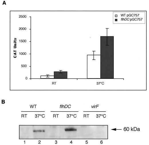
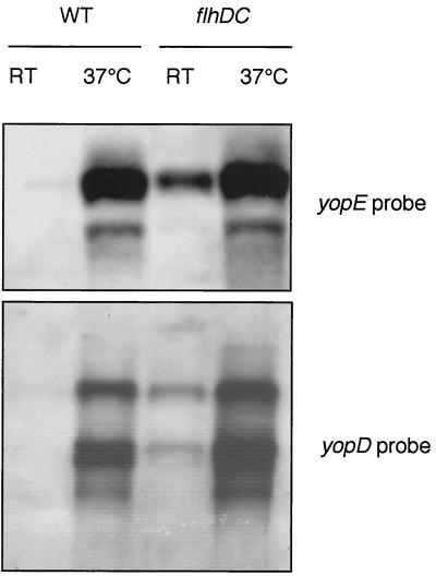
The Yop virulon enables extracellularly located Yersinia, in close contact with a eukaryotic target cell, to inject bacterial toxic proteins directly into the cytosol of this cell. Several Ysc proteins, forming the Yop secretion apparatus, display homology with proteins of the flagellar basal body. To determine whether this relationship could extend to the regulatory pathways, we analyzed the influence of flhDC, the master regulatory operon of the flagellum, on the yop regulon. In an flhDC mutant, the yop regulon was up-regulated. The transcription of virF and the steady-state level of the transcriptional activator VirF were enhanced. yop transcription was increased at 37 degrees C and could also be detected at a low temperature. Yop secretion was increased at 37 degrees C and occurred even at a low temperature. The Ysc secretion machinery was thus functional at room temperature in the absence of flagella, implying that in wild-type bacteria, FlhD and/or FlhC, or the product of a gene downstream of flhDC, represses the yop regulon. In agreement with this notion, increased expression of flhDC in wild-type bacteria resulted in the oversecretion of flagellins at room temperature and in decreased Yop secretion at 37 degrees C.


Figures

FIG. 1.
FIG. 1.
Motility phenotype of the flhDC mutant. (A) Motility assay with a BHI-Ox semisolid plate inoculated with Y. enterocolitica MRS40(pYV40) (wild type [WT]), SBY40(pYV40) (flhDC), and MI1024(pYV1024) (fliA). (B) Western blot analysis with antiflagellin polyclonal antibodies of proteins from whole-cell extracts (C) or from culture supernatants (SN) of Y. enterocolitica MRS40(pYV40) (WT) or SBY40(pYV40) (flhDC) grown for 6 h at RT in BHI-Ox. Lanes were loaded with 8 × 10⁸ bacteria or the supernatant from 10 × 1010 bacteria. The three Y. enterocolitica flagellins are indicated on the left.
FIG. 2.
FIG. 2.
Yop and flagellin secretion by flhDC mutant bacteria and by bacteria overexpressing flhDC. (A) SDS-PAGE and Coomassie brilliant blue staining of proteins from the supernatants of Ca2+-deprived cultures of Y. enterocolitica MRS40(pYV40) (wild type [WT]) (lanes 1 and 5), SBY22703(pSW2276) (yscN flhDC) (lanes 2 and 6), SBY40(pYV40) (flhDC) (lanes 3 and 7), and SBY40(pYV40) with a compensatory mutation (flhDC*) (lanes 4 and 8) after 2 h at RT and 4 h at 37°C (lanes 1 to 4) or 6 h at RT (lanes 5 to 8). The number of bacteria corresponding to the volume of supernatant loaded is given below the panels. Lanes 4 and 8 (flhDC*) were separated from the others because they were not from the same gel. (B) Western blot analysis with antiflagellin polyclonal antibodies of proteins from whole-cell extracts (C) or culture supernatants (SN) of Y. enterocolitica MRS40(pYV40) (WT) or SBY40(pYV40) (flhDC) containing pGY10 or MRS40(pYV40) (WT) containing pGY10 and grown for 6 h at RT in BHI-Ox. Lanes were loaded with 8 × 10⁸ bacteria or the supernatant from 10 × 109 bacteria. (C) SDS-PAGE and Coomassie brilliant blue staining of proteins from the supernatants of Ca2+-deprived cultures of Y. enterocolitica MRS40(pYV40) (WT), SBY40(pYV40) (flhDC) containing pGY10, and MRS40(pYV40) (WT) containing pGY10 after 2 h at RT and 4 h at 37°C. Lanes were loaded with the supernatant from 8 × 10⁹ bacteria. In panels A and C, the positions of the Yops (YopO, -H, -M, -B, -D, -N, -E, and -Q), LcrV (V), and YscP (P) and of proteins that cannot be detected by Coomassie brilliant blue staining (asterisks) are indicated.
FIG. 3.
FIG. 3.
Western blot analysis of intra- and extracellular proteins. Shown is immunodetection (anti-YscJ, -YopN, and -YopE polyclonal antibodies or anti-YopB monoclonal antibody) of proteins from whole-cell extracts (C) (lanes 1, 3, 5, and 7) and culture supernatants (SN) (lanes 2, 4, 6, and 8) of Y. enterocolitica MRS40(pYV40) (wild type [WT]) (lanes 1 to 4) or SBY40(pYV40) (flhDC) (lanes 5 to 8) grown for 6 h at RT (lanes 1, 2, 5, and 6) or 2 h at RT and 4 h at 37°C (lanes 3, 4, 7, and 8) in BHI-Ox. The number of bacteria loaded or the number of bacteria corresponding to the volume of supernatant loaded is given below the panels.
FIG. 4.
FIG. 4.
Other phenotypes of the flhDC mutant. (A) SDS-PAGE and Coomassie brilliant blue staining of proteins from whole-cell extracts (C) and culture supernatants (SN) of Y. enterocolitica MRS40(pYV40) (wild type [WT]) and SBY40(pYV40) (flhDC) grown for 6 h at RT (RT) or 2 h at RT and 4 h at 37°C (37°C) in BHI-Ox. The number of bacteria loaded or the number of bacteria corresponding to the volume of supernatant loaded is given below the panel. (B) Microscopic views (×40) showing total cultures of Y. enterocolitica MRS40(pYV40) (WT) (left panels) or SBY40(pYV40) (flhDC) (right panels) fixed and stained with crystal violet after 6 h at RT (upper panels) or 2 h at RT and 4 h at 37°C (lower panels) in BHI-Ox. (C) Western blot analysis with anti-PhoE polyclonal antibodies of proteins from whole-cell extracts of Y. enterocolitica MRS40(pYV40) (WT) or SBY40(pYV40) (flhDC) grown for 6 h at RT (RT) or 2 h at RT and 4 h at 37°C (37°C) in BHI-Ox. A total of 8 × 10⁸ bacteria were loaded in each lane.
FIG. 5.
FIG. 5.
Effect of the flhDC mutation on VirF. (A) Chloramphenicol acetyltransferase (CAT) activity was measured in extracts of Y. enterocolitica MRS40 (wild type [WT]) and SBY40 (flhDC) cured of the pYV plasmid and carrying pGC757, which encodes a virF-cat operon fusion. Bacteria were grown for 6 h at RT (RT) or 2 h at RT and 4 h at 37°C (37°C) in BHI-Ox. CAT activity was expressed in arbitrary units per unit of optical density of the bacterial suspension at 600 nm. The values represent the averages obtained from four independent experiments, including the standard deviations between the measurements. (B) Western blot analysis with anti-VirF polyclonal antibodies of proteins from whole-cell extracts of Y. enterocolitica MRS40(pYV40) (WT) (lanes 1 and 2), SBY40(pYV40) (flhDC) (lanes 3 and 4), and W22703(pGC1153) (virF) (lanes 5 and 6) grown for 6 h at RT (lanes 1, 3, and 5) or 2 h at RT and 4 h at 37°C (lanes 2, 4, and 6) in BHI-Ox. A total of 8 × 10⁸ bacteria were loaded in each lane.
FIG. 6.
FIG. 6.
Effect of the flhDC mutation on the transcription of yopE and yopD. Shown is Northern blot analysis of yopE and yopD in Y. enterocolitica MRS40(pYV40) (wild type [WT]) or SBY40(pYV40) (flhDC) grown for 4 h at RT (RT) or 2 h at RT and 2 h at 37°C (37°C). The yopE transcript was detected with a PCR product amplified with oligonucleotides MIPA 538 (5′-GCCCCCATGGAAATATCATCATTTATTTCTACAT-3′) and MIPA 539 (5′-CCGGAATTCGCCCCTTGTTTTTATCC-3′), and the yopD transcript was detected with MIPA 829 (5′-CGGGGATCCATGACAATAAATATCAAGACAGAC-3′) and MIPA 830 (5′-CGCGTCGACTCAGACAACACCAAAAGC-3′).
FIG. 7.
FIG. 7.
FlhD and FlhC control the synthesis of the Ysc injectisome. Shown is Western blot analysis of YopE in whole-cell extracts (C) (lanes 1, 3, 5, and 7) and supernatants (SN) (lanes 2, 4, 6, and 8) of Y. enterocolitica MRS40(pAB4052) (yopE) (lanes 1, 2, 5, and 6) and SBY40(pAB4052) (yopE flhDC) (lanes 3, 4, 7, and 8) carrying pSI55 (p lac yopE sycE) and grown for 6 h at RT (lanes 1 to 4) or 2 h at RT and 4 h at 37°C (lanes 5 to 8) in BHI-Ox. The number of bacteria loaded or the number of bacteria corresponding to the volume of supernatant loaded is given below the panel.

References

    1. Bartlett, D. H., B. B. Frantz, and P. Matsumura. 1988. Flagellar transcriptional activators FlbB and FlaI: gene sequences and 5′ consensus sequences of operons under FlbB and FlaI control. J. Bacteriol. 170:1575-1581. - PMC - PubMed
    1. Bertin, P., E. Terao, E. H. Lee, P. Lejeune, C. Colson, A. Danchin, and E. Collatz. 1994. The H-NS protein is involved in the biogenesis of flagella in Escherichia coli. J. Bacteriol. 176:5537-5540. - PMC - PubMed
    1. Bleves, S., and G. R. Cornelis. 2000. How to survive in the host: the Yersinia lesson. Microbes Infect. 2:1451-1460. - PubMed
    1. Blocker, A., P. Gounon, E. Larquet, K. Niebuhr, V. Cabiaux, C. Parsot, and P. Sansonetti. 1999. The tripartite type III secreton of Shigella flexneri inserts IpaB and IpaC into host membranes. J. Cell Biol. 147:683-693. - PMC - PubMed
    1. Cornelis, G., C. Sluiters, C. L. de Rouvroit, and T. Michiels. 1989. Homology between VirF, the transcriptional activator of the Yersinia virulence regulon, and AraC, the Escherichia coli arabinose operon regulator. J. Bacteriol. 171:254-262. - PMC - PubMed

Publication types

MeSH terms

LinkOut - more resources