. 2002 Jun 3;21(11):2557-67.
doi: 10.1093/emboj/21.11.2557.

A tubular EHD1-containing compartment involved in the recycling of major histocompatibility complex class I molecules to the plasma membrane

Affiliations

A tubular EHD1-containing compartment involved in the recycling of major histocompatibility complex class I molecules to the plasma membrane

Steve Caplan et al. EMBO J. .

Abstract

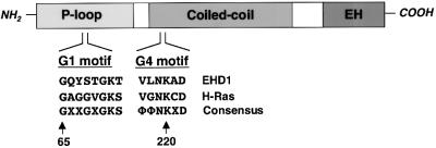
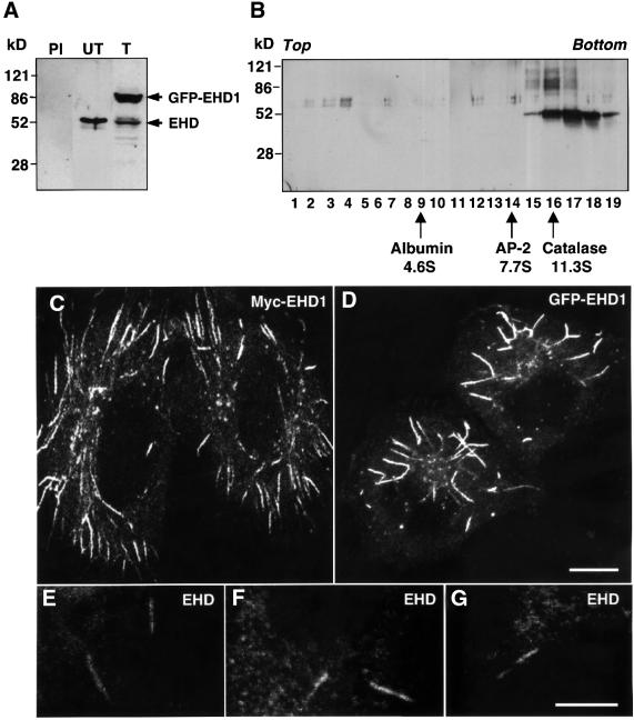
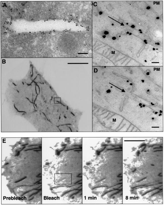
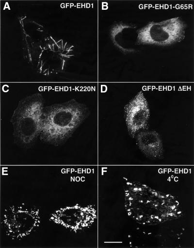
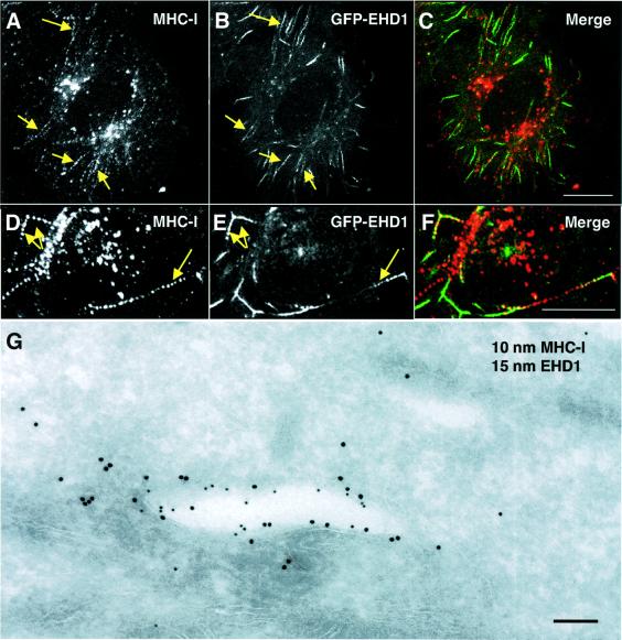
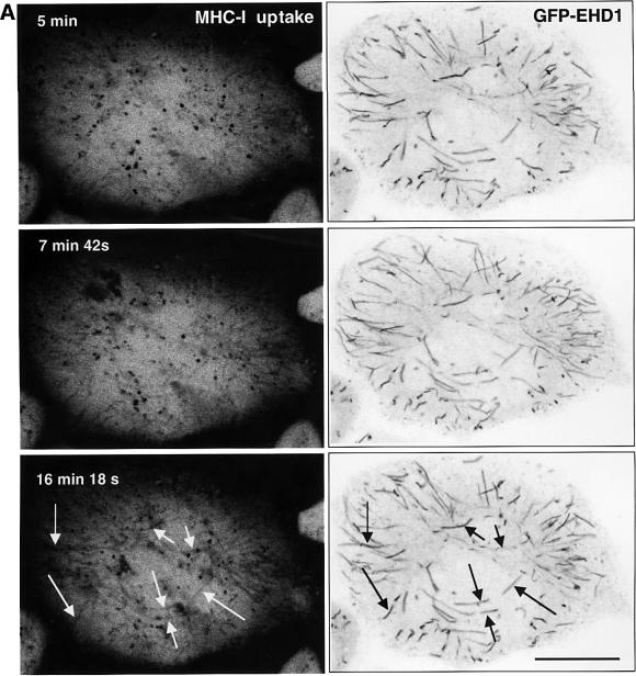
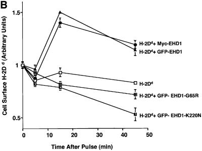
The Eps15 homology (EH) domain-containing protein, EHD1, has recently been ascribed a role in the recycling of receptors internalized by clathrin-mediated endocytosis. A subset of plasma membrane proteins can undergo internalization by a clathrin-independent pathway regulated by the small GTP-binding protein ADP-ribosylation factor 6 (Arf6). Here, we report that endogenous EHD proteins, as well as transgenic tagged EHD1, are associated with long, membrane-bound tubules containing Arf6. EHD1 appears to induce tubule formation, which requires nucleotide cycling on Arf6 and intact microtubules. Mutations in the N-terminal P-loop domain or deletion of the C-terminal EH domain of EHD1 prevent association of EHD1 with tubules or induction of tubule formation. The EHD1 tubules contain internalized major histocompatibility complex class I (MHC-I) molecules that normally traffic through the Arf6 pathway. Recycling assays show that overexpression of EHD1 enhances MHC-I recycling. These observations suggest an additional function of EHD1 as a tubule-inducing factor in the Arf6 pathway for recycling of plasma membrane proteins internalized by clathrin-independent endocytosis.


Figures

None
Fig. 1. EHD1 domain organization and homology to GTP-binding proteins. A schematic representation of human EHD1. EHD1 comprises an N-terminal P-loop, a central coiled coil and a C-terminal EH domain. EHD1 motifs that conform to polypeptide loops involved in GTP binding are shown at amino acids 65–72 (G1) and 217–222 (G4). Note that G2 and G5 motifs (which are more heterogeneous) have not been identified in EHD1, and a sequence with low homology to the G3 motif consensus is found between amino acids 351 and 358 (data not shown). The G1 and G4 amino acid sequences of EHD1 are aligned with those of the GTP-binding protein H-Ras, and with a consensus sequence for Ras-family GTP-binding motifs. X represents any amino acid; Φ represents a bulky hydrophobic amino acid.
None
Fig. 2. EHD1 localizes to an array of long tubular structures. (A) Detergent lysates were prepared from untransfected HeLa cells (PI and UT) or HeLa cells transfected with a GFP–EHD1 construct (T), and resolved by 4–20% SDS–PAGE. Immunoblot analysis with either pre-immune serum (PI) or rabbit polyclonal antibody directed against EHD1 (UT and T) revealed the presence of both endogenous EHD1 (55 kDa) and transgenic GFP–EHD1 (85 kDa) proteins. Following transient transfection, ∼60% of the cells expressed detectable levels of EHD1, and the relative levels of transfected and endogenous proteins were estimated by densitometric analysis of multiple film exposures. (B) HeLa cell extracts were subjected to sedimentation velocity analysis on a 4–20% sucrose gradient. Fractions were collected, resolved by 4–20% SDS–PAGE, and proteins were visualized by immunoblot analysis using the polyclonal antibody prepared against EHD1. Size markers indicate the positions of albumin, AP-2 complex and catalase on the sucrose gradients. (C) HeLa cells were transfected with a plasmid encoding Myc-EHD1. Cells were fixed and permeabilized 24 h later, and incubated with a mouse monoclonal antibody to the Myc epitope. Bound antibodies were revealed by incubation with Cy3-conjugated donkey anti-mouse IgG, demonstrating the presence of a dense network of Myc-EHD1 tubular organelles. (D) HeLa cells were transfected with a plasmid encoding GFP–EHD1, and were fixed and permeabilized after 24 h. (EG) Untransfected HeLa cells were fixed, permeabilized and incubated with a rabbit polyclonal antibody to endogenous EHD1. Bound antibodies were revealed by incubation with Cy3-conjugated donkey anti-rabbit IgG. Images show the presence of long, tubular structures containing endogenous EHD proteins. All images were obtained by confocal microscopy. Bars: (C and D), 10 µm; (E–G), 10 µm.
None
Fig. 3. Ultrastructural analysis of EHD1 tubules. (A) HeLa cells were transiently transfected with Myc-EHD1 and processed for electron microscopy immunogold labeling using an anti-Myc antibody. Bound antibodies were detected using gold-conjugated protein A. (BD) Correlative fluorescence/electron microscopy. HeLa cells were transiently transfected with a GFP–EHD1 construct on CELLocate grids, and live confocal images of a typical GFP–EHD1-expressing cell were obtained (B). Cells were then fixed with 4% paraformaldehyde and 0.05% glutaraldehyde, and enhanced gold labeling was performed for anti-GFP antibodies as described in Materials and methods. (C) and (D) are serial sections depicting the boxed region of interest in (B), and arrows in (C) and (D) mark the EHD1 tubular structure within the box. Particles indicate the presence of GFP–EHD1 along the tubular structure and in the cytosol. (E) Dynamics of EHD1 association with membranes. GFP–EHD1 was subjected to fluorescence recovery after photobleaching (FRAP) analysis. HeLa cells were transiently transfected with a GFP–EHD1 construct, and examined by live confocal image analysis 24 h later. An entire EHD1 tubular structure was photobleached (rectangular region of interest). GFP–EHD1 recovery to the bleached area was monitored every 3.3 s (see Supplementary time-lapse video). Images of live cells (B and E) are visualized as inverted images to facilitate analysis. M, mitochondria; PM, plasma membrane. Bars: (A), 200 nm; (B), 10 µm; (C and D), 200 nm; (E), 10 µm.
None
Fig. 4. Requirements for EHD1 tubule formation. HeLa cells were transiently transfected with cDNA constructs coding for wild-type GFP–EHD1 (A), GFP–EHD1-G65R (B), GFP–EHD1-K220N (C), or GFP–EHD1 ΔEH (D), fixed 24 h later, and analyzed by confocal microscopy. Wild-type GFP–EHD1-transfected HeLa cells were also treated for 1.5 h with nocodazole (NOC) (E), or maintained at 4°C for 1 h (F) prior to fixation. Images were obtained by confocal microscopy. Bar, 10 µm.
None
Fig. 5. Co-localization and functional interaction of EHD1 with Arf6. HeLa cells were transiently co-transfected with constructs encoding Myc-EHD1 and wild-type Arf6 (AC), GFP–EHD1 and Arf6-Q67L (DF), GFP–EHD1 and Arf6-T27N (GI), GFP–EHD1 and FLAG-EFA6 (JL), and GFP–EHD1 and FLAG-ACAP1 (MO). Cells were fixed, permeabilized and incubated with a monoclonal antibody to the Myc epitope and a rabbit polyclonal antibody to Arf6 (A–C). Bound antibodies were revealed by Alexa-488-conjugated antibody to mouse IgG (A and C), and by Cy3-conjugated anti-rabbit IgG (B and C). HeLa cells co-transfected with GFP–EHD1 and Arf6 mutant constructs (D–I) were fixed, permeabilized and incubated with a rabbit polyclonal antibody directed against Arf6 (D–I), followed by Cy3-conjugated anti-rabbit IgG (E, F, H and I). HeLa cells co-transfected with GFP–EHD1 and FLAG-EFA6 (J–L) or FLAG-ACAP1 (M–O) were fixed, permeabilized and incubated with a monoclonal antibody to the FLAG epitope, followed by a Cy3-conjugated anti-mouse IgG antibody. All images were obtained by confocal microscopy. Arrows (A and B) denote tubular structures containing both Arf6 and EHD1. Bar, 10 µm.
None
Fig. 6. Co-localization of EHD1 and internalized MHC-I molecules. (AF) Co-localization of internalized MHC-I with EHD1 tubular structures. HeLa cells were transiently transfected with a construct encoding GFP–EHD1. After 24 h, the cells were continuously pulsed with W6/32 anti-MHC-I monoclonal antibody for 30 min. After brief acid washing to remove surface-bound MHC-I antibody, fixed and permeabilized cells were incubated with Cy3-conjugated anti-mouse IgG, and examined by confocal microscopy. Internalized MHC-I is shown in (A) and (D), and GFP–EHD1 is depicted in (B) and (E). (C and F) Merged images. (G) HeLa cells were transiently co-transfected with Myc-EHD1 and H-2Dd (mouse MHC-I) constructs. Cells were pulsed with anti-H-2Dd antibody 24 h later, fixed and processed for ultrathin section electron microscopy. EHD1 is marked by 15 nm gold particles, and 10 nm gold particles mark the presence of internalized H-2Dd. Arrows denote tubular structures positive for internalized MHC-I (A and D) and GFP–EHD1 (B and E). Bars: (A–C), 10 µm; (D–F), 10 µm; (G), 200 nm.
None
Fig. 7. EHD1 tubular structures promote recycling of MHC-I to the cell surface. (A) Time-dependent co-localization of internalized MHC-I with EHD1 tubules by live image analysis. MHC-I monoclonal antibodies were coupled to Alexa Fluor 568 F(ab′)2 fragment of goat anti-mouse IgG. The coupled antibodies were then used to continuously pulse HeLa cells that were transfected 24 h earlier with a GFP–EHD1 construct. Images of MHC-I uptake (left panels) and GFP–EHD1 tubules (right panels) are depicted. Arrows (white) mark MHC-I tubular structures that appear at 15–20 min of internalization and co-localize with pre-existing GFP–EHD1 tubules (black arrows). Images are shown inverted to facilitate analysis (see Supplementary time-lapse video). Bar, 10 µm. (B) Quantification of EHD1-enhanced MHC-I recycling by a CELISA assay. HeLa cells were transfected with cDNA coding for H-2Dd (mouse MHC-I), H-2Dd and GFP–EHD1, H-2Dd and Myc-EHD1, H-2Dd and GFP–EHD1-G65R, or H-2Dd and GFP–EHD1-K220N. Internalization of MHC-I over time was monitored 24 h after transfection by CELISA utilizing a biotinylated anti-MHC-I antibody (see Materials and methods), and the fraction of MHC-I antibody on the surface at each time point was recorded. A representative experiment from four independent CELISA assays is depicted, with triplicates at each time point.

References

    1. Abdel-Motal U.M., Berg,L., Bengtsson,M., Dahmen,J., Kihlberg,J., Magnusson,G., Nilsson,U. and Jondal,M. (1995) Major histocompatibility complex class I binding glycopeptides for the estimation of ‘empty’ class I molecules. J. Immunol. Methods, 188, 21–31. - PubMed
    1. Bonifacino J.S. and Dell’Angelica,E.C. (1999) Molecular bases for the recognition of tyrosine-based sorting signals. J. Cell Biol., 145, 923–926. - PMC - PubMed
    1. Brodsky F.M., Chen,C.Y., Knuehl,C., Towler,M.C. and Wakeham,D.E. (2001) Biological basket weaving: formation and function of clathrin-coated vesicles. Annu. Rev. Cell Dev. Biol., 17, 517–568. - PubMed
    1. Brown F.D., Rozelle,A.L., Yin,H.L., Balla,T. and Donaldson,J.G. (2001) Phosphatidylinositol 4,5-bisphosphate and Arf6-regulated membrane traffic. J. Cell Biol., 154, 1007–1017. - PMC - PubMed
    1. Caplan S., Dell’Angelica,E.C., Gahl,W.A. and Bonifacino,J.S. (2000) Trafficking of major histocompatibility complex class II molecules in human B-lymphoblasts deficient in the AP-3 adaptor complex. Immunol. Lett., 72, 113–117. - PubMed

Publication types

MeSH terms