. 2002 Jun 3;21(11):2703-14.
doi: 10.1093/emboj/21.11.2703.

Microphthalmia-associated transcription factor interacts with LEF-1, a mediator of Wnt signaling

Affiliations

Microphthalmia-associated transcription factor interacts with LEF-1, a mediator of Wnt signaling

Ken-ichi Yasumoto et al. EMBO J. .

Abstract

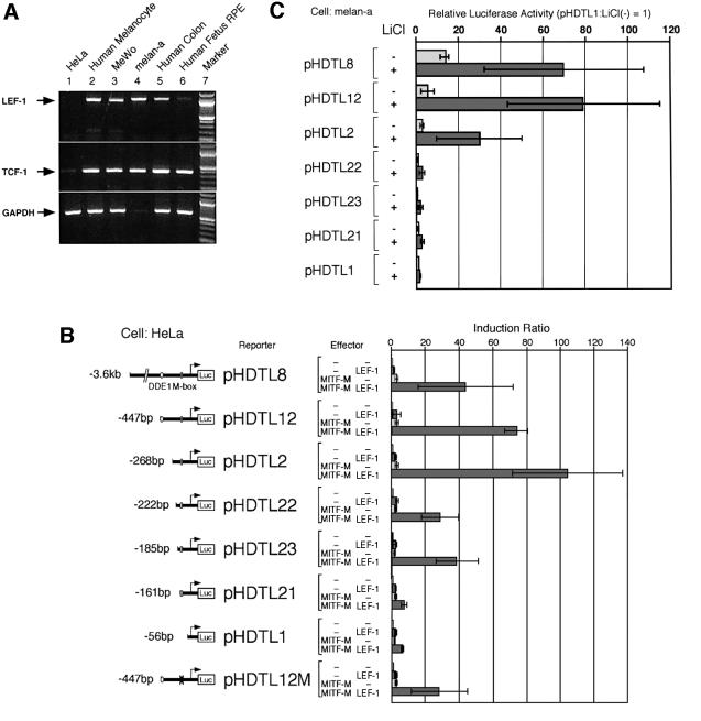
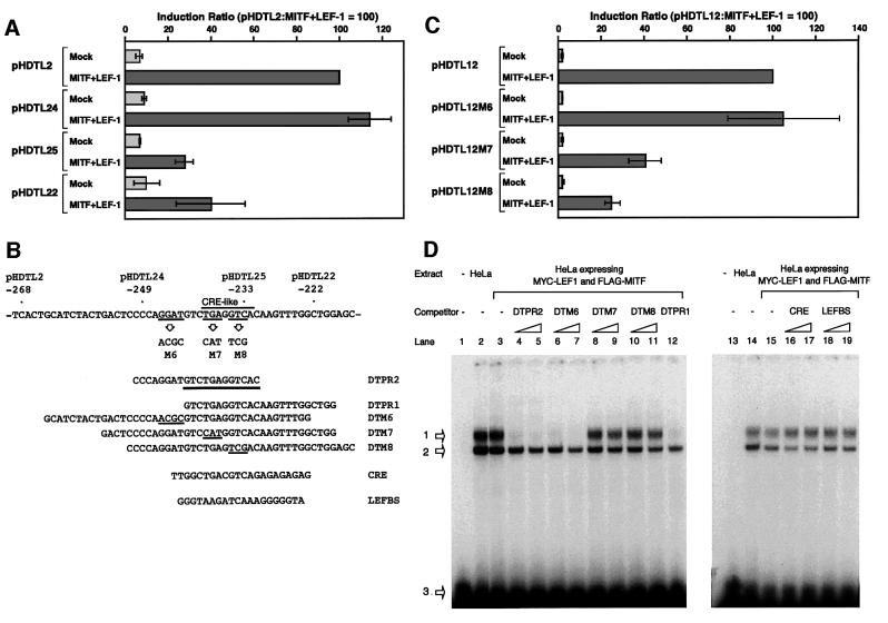
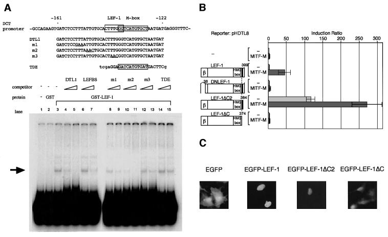
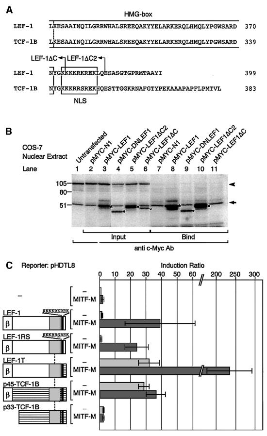
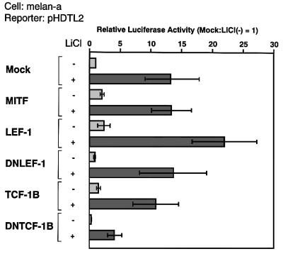
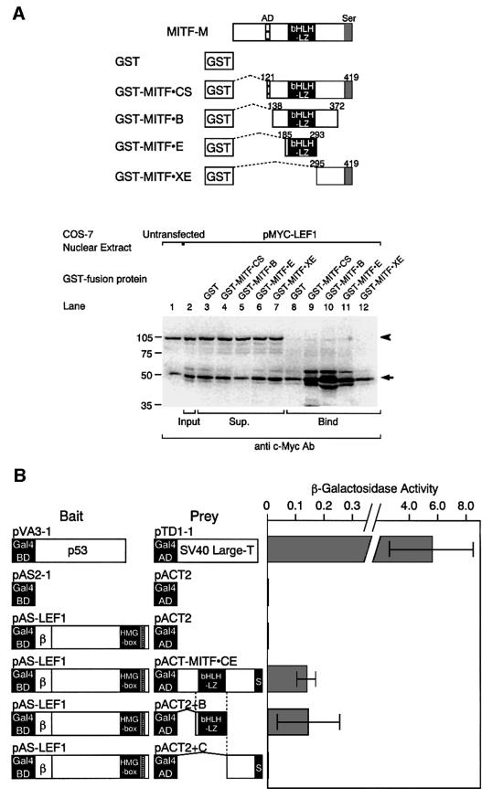
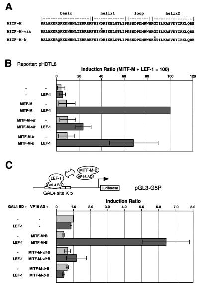
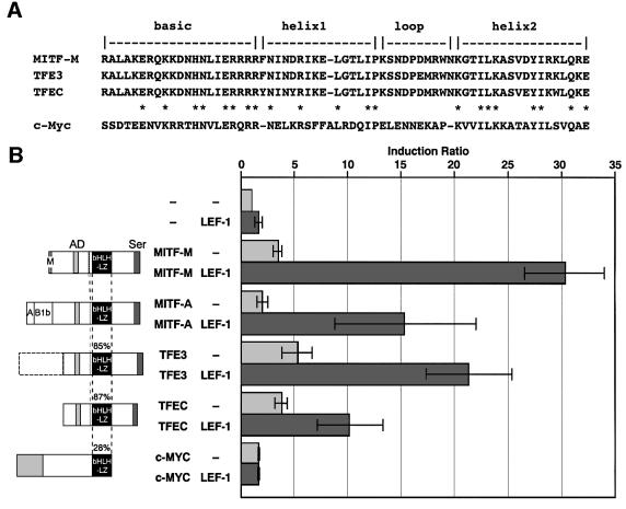
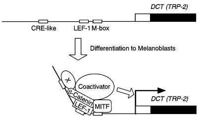
Wnt signals regulate differentiation of neural crest cells through the beta-catenin associated with a nuclear mediator of the lymphoid-enhancing factor 1 (LEF-1)/T-cell factors (TCFs) family. Here we show the interaction between the basic helix-loop-helix and leucine-zipper region of microphthalmia-associated transcription factor (MITF) and LEF-1. MITF is essential for melanocyte differentiation and its heterozygous mutations cause auditory-pigmentary syndromes. Functional cooperation of MITF with LEF-1 results in synergistic transactivation of the dopachrome tautomerase (DCT) gene promoter, an early melanoblast marker. This activation depends on the separate cis-acting elements, which are also responsible for the induction of the DCT promoter by lithium chloride that mimics Wnt signaling. beta-catenin is required for efficient transactivation, but dispensable for the interaction between MITF and LEF-1. The interaction with MITF is unique to LEF-1 and not detectable with TCF-1. LEF-1 also cooperates with the MITF-related proteins, such as TFE3, to transactivate the DCT promoter. This study therefore suggests that the MITF/TFE3 family is a new class of nuclear modulators for LEF-1, which may ensure efficient propagation of Wnt signals in many types of cells.


Figures

None
Fig. 1. Effects of LEF-1 on the DCT promoter activity. (A) Expression profiles of LEF-1 and TCF-1 mRNAs determined by RT–PCR. Note the faint band of GAPDH in mouse melan-a cells. (B) Promoter-context dependent transactivation of the DCT promoter by MITF-M and LEF-1. Each of the DCT reporter plasmids was coexpressed in HeLa cells with MITF-M, LEF-1 or a combination of MITF-M and LEF-1. An enhancer DDE1 (positions –447 to –416) and the M box (positions –138 to –128) are indicated. The magnitude of activation is presented as the ratio of normalized luciferase activity obtained with each plasmid and that with vector DNA (Induction Ratio). The results of at least three independent experiments are shown with standard deviations. (C) Activation of DCT promoter by LiCl. Melan-a cells, maintained in a 6-well plate, were transfected with the indicated constructs (1 µg each of reporter and effector and 0.05 µg of pCDNA3-His-LacZ), incubated for 20 h, and then treated with 30 mM LiCl for 24 h in fresh medium. The data are shown as a ratio to the basal luciferase activity obtained with pHDTL1. Other conditions were the same as in (B).
None
Fig. 2. Identification of the cis-acting element that is required for the activation of DCT promoter by LEF-1 and MITF-M. (A) Deletion studies of the DCT promoter. HeLa cells were cotransfected with the indicated reporter plasmids. (B) The cis-acting region in the DCT promoter. The 12-bp cis-acting element is underlined in DTPR2. Also shown is the strategy for the functional analysis in HeLa cells (C) and EMSAs (D). Base changes shown were introduced into construct pHDTL12. Nuclear extracts of HeLa cells were incubated with a 32P-end labeled DTPR2 in the absence (lanes 2, 3, 14 and 15) or presence of an indicated competitor (200- and 500-fold excesses, shown as triangles). LEFBS represents a LEF-1-binding site. Lanes 1 and 13 represent a control lacking nuclear extracts. Arrows 1 and 2 indicate the specific and unspecific protein–DNA complexes, respectively. Unbound probes are indicated by arrow 3.
None
Fig. 3. Functional cooperation of MITF-M with LEF-1. (A) EMSAs showing the LEF-1-binding site of the DCT promoter. GST–LEF-1 fusion protein was incubated with a 32P-end labeled DTL1 in the absence (lane 3) or presence of indicated competitors (200- and 500-fold excesses, shown as triangles). Lanes 1 and 2 represent a buffer control and GST control. TDE represents the distal enhancer of the tyrosinase gene that is bound by MITF-M (Yasumoto et al., 1994). An arrow indicates the specific protein–DNA complex. (B) Domains of LEF-1 required for the DCT promoter activation. HeLa cells were cotransfected with a DCT reporter plasmid, pHDTL8, and indicated LEF-1 plasmids. Shown are the N-terminal β-catenin-binding domain (β) and the NLS near the C-terminus (closed box). Reporter luciferase activity obtained was normalized with each β-galactosidase activity that represents an internal control. The magnitude of activation is presented as the ratio of normalized luciferase activity obtained with each effector plasmid and that with vector DNA. (C) Subcellular localization of EGFP–LEF-1 fusion proteins in COS-7 cells, as assessed by fluorescence.
None
Fig. 4. Interaction of MITF-M with the C-terminal region of LEF-1. (A) Alignment of the C-termini. Broken line represents the position where LEF-1 cDNA was replaced by TCF-1B cDNA. (B) Western blot analysis of the c-Myc-tagged LEF-1 proteins bound to MITF-M. Tagged LEF-1 (asterisks) comigrated with endogenous c-Myc, present in COS-7 nuclear extracts (lanes 1–11), indicated by an arrow. The unspecific signal of 105 kDa is indicated by an arrowhead (lanes 1–6). Mutant LEF-1 proteins are shown by closed circles and LEF-1ΔC, an open circle. (C) Functions of LEF-1–TCF-1 chimeric proteins in HeLa cells. Other conditions were the same as in Figure 2A.
None
Fig. 5. Functional difference between LEF-1 and TCF-1B. Melan-a cells were transfected with pHDTL2 and the indicated constructs, and then treated for 24 h with 30 mM LiCl. Other conditions were the same as in Figure 1C.
None
Fig. 6. Interaction of the bHLH/LZ region of MITF-M with LEF-1. (A) In vitro interaction between LEF-1 and the bHLH/LZ domain of MITF. COS-7 nuclear extracts contained endogenous c-Myc (lanes 1–12), as indicated by an arrow. Tagged LEF-1 was detected as enhanced signals in the fractions, bound to truncated MITF-M proteins containing the bHLH/LZ domain (lanes 9–11). An arrowhead indicates the unspecific protein binding (lanes 1–7). (B) Interaction between LEF-1 and the bHLH/LZ region of MITF-M in yeast cells.
None
Fig. 7. Identification of a crucial amino acid in the bHLH/LZ region of MITF-M for protein interaction. (A) The amino acid substitutions in the bHLH/LZ region (asterisks). (B) Effects on functional cooperation. HeLa cells were cotransfected with a DCT-luciferase reporter plasmid (pHDTL8), LEF-1 and the indicated MITF-M proteins. The results of three independent experiments are shown with standard deviations. (C) Effects on physical interaction. Two-hybrid assays were performed in 293 human embryonic kidney cells using the fusion proteins containing a bHLH/LZ region of MITF-M or its mutant proteins.
None
Fig. 8. Functional cooperation of LEF-1 with MITF-related proteins. (A) Comparison of the bHLH/LZ regions. (B) Cooperation of LEF-1 with various bHLH/LZ proteins. The amino acid identity is shown as a percentage of the MITF bHLH/LZ region. HeLa cells were cotransfected with pHDTL8 and the indicated combination of LEF-1 and various bHLH/LZ proteins. An equal amount of MITF constructs was used (2 µg each), and the total amounts of plasmid DNA were maintained at 4 µg with the vector DNA (pRc/CMV). The data are presented as the ratio of normalized luciferase activity obtained with each combination and that obtained with vector DNA.
None
Fig. 9. Proposed model for transcriptional activation of the DCT gene. The relevant cis-acting elements are shown schematically. Note that a CRE-like motif-binding protein (X) is endogenously expressed in HeLa cells and melanoma cells (see Figure 2D and the relevant text).

References

    1. Amae S. et al. (1998) Identification of a novel isoform of microphthalmia-associated transcription factor that is enriched in retinal pigment epithelium. Biochem. Biophys. Res. Commun., 247, 710–715. - PubMed
    1. Amae S., Yasumoto,K., Takeda,K., Udono,T., Takahashi,K. and Shibahara,S. (2000) Identification of a composite enhancer of the human tyrosinase-related protein 2/DOPAchrome tautomerase gene. Biochim. Biophys. Acta, 1492, 505–508. - PubMed
    1. Amiel J., Watkin,P.M., Tassabehji,M., Read,A.P. and Winter,R.M. (1998) Mutation of the MITF gene in albinism–deafness syndrome (Tietz syndrome). Clin. Dysmorphol., 7, 17–20. - PubMed
    1. Atchley W.R. and Fitch,W.M. (1997) A natural classification of the basic helix–loop–helix class of transcription factors. Proc. Natl Acad. Sci. USA, 94, 5172–5176. - PMC - PubMed
    1. Barker N., Morin,P.J. and Clevers,H. (2000) The yin-yang of TCF/ β-catenin signaling. Adv. Cancer Res., 77, 1–24. - PubMed

Publication types

MeSH terms

LinkOut - more resources