. 2002 May 28;99(11):7402-7.
doi: 10.1073/pnas.072199199.

Amyloid aggregates of the HET-s prion protein are infectious

Affiliations

Amyloid aggregates of the HET-s prion protein are infectious

Marie-Lise Maddelein et al. Proc Natl Acad Sci U S A. .

Abstract

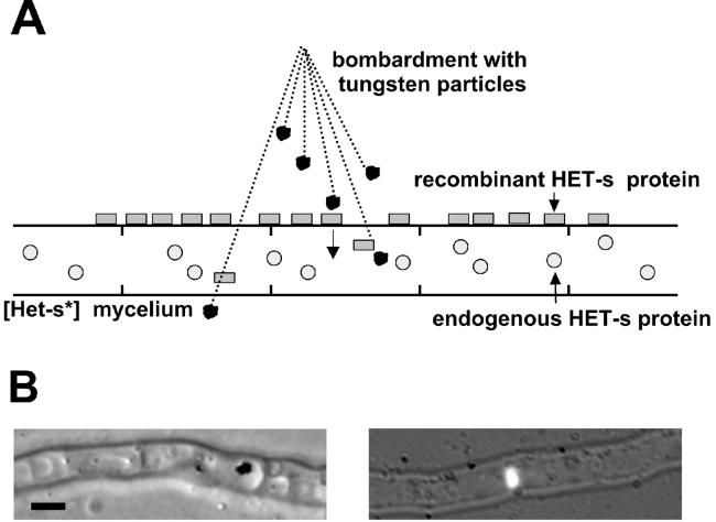
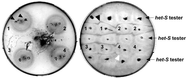
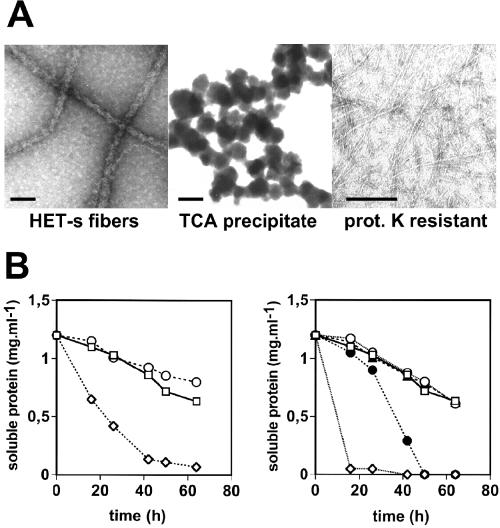
The [Het-s] infectious element of the filamentous fungus Podospora anserina is a prion. We have recently reported that recombinant HET-s protein aggregates in vitro into amyloid fibers. In vivo, the protein aggregates specifically in the [Het-s] prion strains. Here, we show that biolistic introduction of aggregated recombinant HET-s protein into fungal cells induces emergence of the [Het-s] prion with a high frequency. Thus, we demonstrate that prion infectivity can be created de novo, in vitro from recombinant protein in this system. Although the amyloid filaments formed from HET-s could transmit [Het-s] efficiently, neither the soluble form of the protein nor amorphous aggregates would do so. In addition, we have found that (i) [Het-s] infectivity correlates with the ability to convert HET-s to amyloids in vitro, (ii) [Het-s] infectivity is resistant to proteinase K digestion, and (iii) HET-s aggregates formed in vivo in [Het-s] strains have the ability to convert the recombinant protein to aggregates. Together, our data designate the HET-s amyloids as the molecular basis of [Het-s] prion propagation.


Figures

Figure 1
Figure 1
Biolistic introduction of recombinant HET-s protein into [Het-s*] mycelium. (A) Schematic representation of the biolistic assay. Mycelium from a [Het-s*] (prion-free) strain is overlaid with recombinant HET-s protein and bombarded with tungsten particles. (B) Light micrographs of mycelium after bombardment. (Left) Tungsten particle located within a fungal vacuole. (Right) Fluorescein-labeled HET-s aggregates detected within a fungal cell. (Bar = 2 μm.)
Figure 2
Figure 2
Phenotypic detection of the [Het-s] prion after microprojectile bombardment. Petri dish after bombardment (Left). The tungsten particles appear clustered in one spot but are actually dispersed over the entire Petri dish. Two inoculates are sampled from each strain (Left, arrowheads) and subcultured in confrontation with a [Het-S] tester strain for incompatibility tests (Right). The black arrow indicates a cell death reaction (“barrage”), and the white arrow indicates a normal contact line. In this example, strains 1–3 have acquired the [Het-s] prion whereas strain 4 has remained in the [Het-s*] (prion-free) state.
Figure 3
Figure 3
Electron micrographs of HET-s aggregates used in the biolistic assay and their ability to convert recombinant HET-s. (A) HET-s aggregates were prepared and analyzed by electron microscopy after negative staining. (Bar = 50 nm.) (B) In vitro aggregation assays of recombinant soluble HET-s were inoculated in a 1:10 ratio with various HET-s samples. (Left) Control (○); 100,000 × g supernatant of spontaneously aggregating HET-s (□); 100,000 × g pellet of spontaneously aggregating HET-s (◊). (Right) Control (○); trichloroacetic acid (TCA)-precipitated HET-s (□); heat-denatured HET-s (▴); proteinase K-digested HET-s fibers (●); sonicated HET-s fibers (◊). Aggregation assays were performed as described.
Figure 4
Figure 4
Extracts from a [Het-s] strain containing HET-s aggregates convert soluble recombinant HET-s into aggregates. Recombinant soluble HET-s protein (1 mg⋅ml−1) was inoculated with an equal amount (wt/wt) of a sucrose pellet fraction from a [Het-s] strain containing HET-s aggregates ([Het-s] lane), or from the het-s-knockout strain (Δhet-s lane), or with buffer A alone (“no extract” lane) and kept at 4°C. The reaction mixture was centrifuged at 10,000 × g for 15 min after 30 min, 12 h, and 26 h, and supernatant and pellet fractions were analyzed by SDS/PAGE and Coomassie blue staining. The position of recombinant HET-s protein, which migrates at 32 kDa, is marked.

Comment in

References

    1. Prusiner S B. Proc Natl Acad Sci USA. 1998;95:13363–13383. - PMC - PubMed
    1. Kocisko D A, Come J H, Priola S A, Chesebro B, Raymond G J, Lansbury P T, Caughey B. Nature (London) 1994;370:471–474. - PubMed
    1. Kaneko K, Peretz D, Pan K M, Blochberger T C, Wille H, Gabizon R, Griffith O H, Cohen F E, Baldwin M A, Prusiner S B. Proc Natl Acad Sci USA. 1995;92:11160–11164. - PMC - PubMed
    1. Hill A F, Antoniou M, Collinge J. J Gen Virol. 1999;80:11–14. - PubMed
    1. Wickner R B. Science. 1994;264:566–569. - PubMed

Publication types

LinkOut - more resources