. 2002 May 28;99(11):7461-6.
doi: 10.1073/pnas.102596199.

Subunit composition of mammalian transient receptor potential channels in living cells

Affiliations

Subunit composition of mammalian transient receptor potential channels in living cells

Thomas Hofmann et al. Proc Natl Acad Sci U S A. .

Abstract

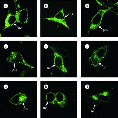
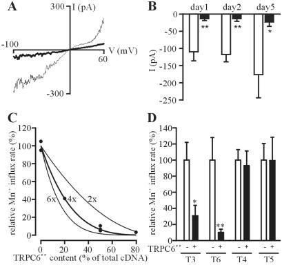
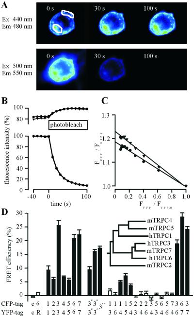
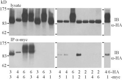
Hormones, neurotransmitters, and growth factors give rise to calcium entry via receptor-activated cation channels that are activated downstream of phospholipase C activity. Members of the transient receptor potential channel (TRPC) family have been characterized as molecular substrates mediating receptor-activated cation influx. TRPC channels are assumed to be composed of multiple TRPC proteins. However, the cellular principles governing the assembly of TRPC proteins into homo- or heteromeric ion channels still remain elusive. By pursuing four independent experimental approaches--i.e., subcellular cotrafficking of TRPC subunits, differential functional suppression by dominant-negative subunits, fluorescence resonance energy transfer between labeled TRPC subunits, and coimmunoprecipitation--we investigate the combinatorial rules of TRPC assembly. Our data show that (i) TRPC2 does not interact with any known TRPC protein and (ii) TRPC1 has the ability to form channel complexes together with TRPC4 and TRPC5. (iii) All other TRPCs exclusively assemble into homo- or heterotetramers within the confines of TRPC subfamilies--e.g., TRPC4/5 or TRPC3/6/7. The principles of TRPC channel formation offer the conceptual framework to assess the physiological role of distinct TRPC proteins in living cells.


Figures

Figure 1
Figure 1
Co-trafficking of TRPC1 and TRPC6Δ131 by members of the TRPC family. C-terminally YFP-tagged hTRPC1A was expressed in HEK293 cells alone (a) or together with untagged hTRPC3 (b) or mTRPC4β (c), and the cellular localization was assessed by confocal laser-scanning microscopy. d shows a cell expressing TRPC6-GFP. An N-terminally truncated TRPC6 mutant C-terminally tagged with GFP (TRPC6Δ131-GFP) was expressed alone (e) or together with untagged hTRPC3 (f), hTRPC6 (g), mTRPC4β (h), or mTRPC5 (i). Typical examples from three independent transfections are shown. ne, nuclear envelope; pm, plasma membrane.
Figure 2
Figure 2
Analysis of TRPC multimerization properties with a dominant-negative TRPC6 mutant (TRPC6DN). (A) Current-voltage relationships of AlFformula image-induced TRPC6 currents in HEK293 cells stably expressing wild-type hTRPC6 (thin trace) and additionally expressing TRPC6DN (bold trace) were recorded. (B) Means ± SEM (n = 6) of peak TRPC6 whole-cell currents at −60 mV holding potential from 6 experiments each with (filled bars) or without (open bars) TRPC6DN coexpressed are shown at different time points after transfection of TRPC6DN-cDNA. Statistical significance is indicated (*, P < 0.05; **, P < 0.01). (C) CHO-K1 cells were comicroinjected with a fixed amount of wild-type TRPC6 and variable amounts of TRPC6DN cDNA as indicated on the abscissa. The rate of receptor-activated manganese influx relative to cells expressing only wild-type TRPC6 is plotted as a function of the relative content of TRPC6DN in the cDNA mixture. Each point represents the mean of 1 independent Mn2+ quenching experiment. The extent of suppression expected for the indicated levels of cooperativity are represented. (D) The relative suppression of Mn2+ influx rates (mean ± SEM of n = 5 independent experiments) in CHO-K1 cells microinjected with cDNAs encoding the wild-type TRPC proteins and equal amounts of TRPC6DN cDNA (filled bars) or vector control (open bars) is depicted. Statistical significance is indicated (*, P < 0.05; **, P < 0.01).
Figure 3
Figure 3
Determination of FRET between TRPC channel subunits. (A) Fluorescence of a cell coexpressing TRPC3-CFP and TRPC6-YFP. The images represent CFP (Upper) or YFP (Lower) fluorescences at different time points during photobleaching of YFP. (B) Time courses of the relative CFP and YFP fluorescence of regions of interest defined in A are shown. Circles represent intracellular fluorescence, and triangles denote fluorescence signals over the plasma membrane. (C) Kinetic correlation of the relative amount of YFP photobleaching and the concomitant increase in CFP fluorescence in the same cell. An extrapolation of the linear regression to FYFP = 0 indicates the actual extent of CFP fluorescence recovery. (D) Different combinations of TRPC channels tagged with either CFP or YFP were coexpressed in HEK293 cells as indicated on the abscissa, and the recovery of CFP fluorescence (expressed as percentage of final CFP fluorescence) during YFP photobleaching was quantified. Each bar represents means ± SEM of at least 3 independent cotransfection experiments. Expression of soluble CFP and YFP served as controls (c), and combinations showing a significant FRET signal (P < 0.05) are indicated by filled bars. The phylogenetic relationships within the TRPC channel family are given (Inset).
Figure 4
Figure 4
Co-immunoprecipitation of TRPC channel subunits. TRPC channels were C-terminally tagged with an HA tag or an myc-tag and coexpressed in HEK293 cells in the combinations indicated below the panels. Anti-HA immunoreactivity was detected by immunoblotting (IB) before (Upper, lysate) or after immunoprecipitation with an anti-myc Ab (Lower, IP-myc).

References

    1. Berridge M J. Neuron. 1998;21:13–26. - PubMed
    1. Montell C, Rubin M R. Neuron. 1989;2:1313–1323. - PubMed
    1. Wes P, Chevesich J, Jeromin A, Rosenberg C, Stetten G, Montell C. Proc Natl Acad Sci USA. 1995;92:9652–9656. - PMC - PubMed
    1. Zhu X, Jiang M, Peyton M, Boulay G, Hurst R, Stefani E, Birnbaumer L. Cell. 1996;85:661–671. - PubMed
    1. Philipp S, Cavalie A, Freichel M, Wissenbach U, Zimmer S, Trost C, Marquart A, Murakami M, Flockerzi V. EMBO J. 1996;15:6166–6171. - PMC - PubMed

Publication types

MeSH terms