. 2002 May 28;99(11):7699-704.
doi: 10.1073/pnas.112012499.

Neuronal deactivation explains decreased cerebellar blood flow in response to focal cerebral ischemia or suppressed neocortical function

Affiliations

Neuronal deactivation explains decreased cerebellar blood flow in response to focal cerebral ischemia or suppressed neocortical function

Lorenz Gold et al. Proc Natl Acad Sci U S A. .

Abstract

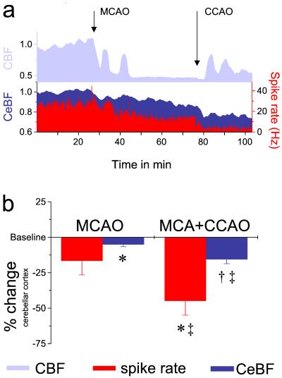
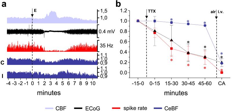
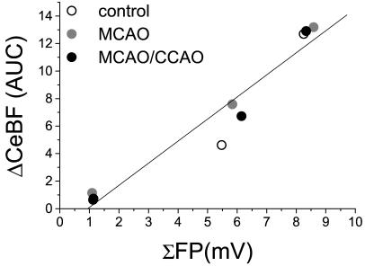
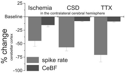
Functional neuroimaging in humans with acute brain damage often reveals decreases in blood flow and metabolism in areas unaffected by the lesion. This phenomenon, termed diaschisis, is presumably caused by disruption of afferent excitatory input from the lesioned area to other brain regions. By characterizing its neurophysiological basis, we used cerebellar diaschisis to study the relationship between electrical activity and blood flow during decreased neuronal activity. Here we show that focal cerebral ischemia in rats causes diaschisis in the cerebellar cortex characterized by pronounced decreases in Purkinje cell spiking activity and small decreases in cerebellar blood flow. The findings were explained by decreased excitatory input to the cerebellar cortex, i.e., deactivation, as cerebellar neuronal excitability and vascular reactivity were preserved. Functional ablation of the cerebral cortex by either spreading depression or tetrodotoxin reproduced the changes in cerebellar function with complete recovery of Purkinje cell activity and cerebellar blood flow concomitant with recovery of neocortical function. Decreases of activity involving the contralateral frontal cortex produced the largest decrease in cerebellar electrical activity and blood flow. Our data suggest that deactivation explains the decreases in blood flow and metabolism in cerebellar diaschisis observed in human neuroimaging studies. Decreases in spiking activity were 3-7 times larger than the respective decreases in flow. Therefore, under pathological conditions, neuroimaging methods based on hemodynamic signals may only show small changes, although the underlying decrease in neuronal activity is much larger.


Figures

Figure 1
Figure 1
Occluding the left middle cerebral artery, either alone or in combination with the common carotid artery, reduces spike activity and blood flow in the contralateral cerebellar hemisphere. (a) Representative data from one experiment: The CBF in the left cerebral hemisphere decreased markedly after MCAO. Transient increases in CBF indicated peri-infarct depolarizations (PIDs). Additional CCAO further lowered CBF and induced repetitive PIDs. MCAO and CCAO induced gradual reductions of Purkinje cell spike rate. Decreases in neuronal activity were reflected by moderate decreases in CeBF. The y axis without units indicates normalized data. (b) Statistical analysis for all ischemia experiments presented as normalized data (n = 10). Data significantly different from preocclusion baseline: *, P < 0.01; †, P < 0.001. Data significantly different from levels after MCAO alone: ‡, P < 0.01.
Figure 2
Figure 2
Unilateral functional ablation of the cerebral cortex induces crossed cerebellar diaschisis. (a) Original recordings from one animal. Cortical spreading depression was elicited (E) in the left parieto-occipital cortex and then reached the site of CBF- and ECoG-recordings in the parieto-frontal, as indicated by the characteristic increase in CBF and the suppression of the ECoG. At the same time, the spontaneous spiking (red) in Crus II of the contralateral cerebellar cortex and both contralateral (C) and ipsilateral CeBF (I) decreased as well. The y axis without units indicates normalized data. (b) Functional ablation of the cerebral cortex with TTX induces crossed cerebellar diaschisis (n = 4). Normalized mean data of 15 min periods of ECoG, as well as spike rate and CeBF in the contralateral cerebellum at baseline and during the 60 min after application of TTX to the left anterior cerebral cortex. The background values obtained 2 min after cardiac arrest (CA) are shown for comparison. *, Values significantly different from baseline (P < 0.05).
Figure 3
Figure 3
Crossed cerebellar diaschisis is best explained by functional ablation of the frontal cortex. (a) Summary of nine CSDs in three animals: CSD was elicited (E) in the left parieto-occipital (n = 5) or frontal cortex (n = 4), propagated over the hemisphere, and subsequently reached ECoG-recordings, in the left parietal (P) and frontal cortex (F), depending on the point of elicitation. (b) Typical example of one CSD, elicited (E) in the left parieto-occipital cortex: Spike rate and CeBF in the contralateral cerebellar hemisphere reached their minimum when CSD-induced suppression of ECoG-activity involved the frontal cortex (F) and the parietal ECoG (P) had already partly recovered. The y axis without units indicates normalized data. Levels of significance compared with control (baseline): *, P < 0.05; †, P < 0.01; ‡, P < 0.001; §, P < 0.0001; ¶, P < 10−5; ∥, P < 10−6. Levels of significance compared with values at maximal suppression of activity in the parietal cortex: **, P < 0.05; ‡‡, P < 0.01.
Figure 4
Figure 4
Coupling between increased electrical activity and blood flow is preserved during cerebellar diaschisis induced by focal cerebral ischemia (n = 3). The graph shows the correlation between the total evoked electrical activity (calculated as the summed field potential; ΣFP) and the coupled CeBF-increase (ΔCeBF; area under curve) evoked by electrical stimulation of the inferior olive at 1, 5, and 10 Hz. The linear correlation (R = 0.97; P < 0.001) was preserved between ΣFP and ΔCeBF before arterial occlusion (control, open circle), after contralateral MCAO (gray circle), and after additional MCAO/CCAO (black circle).
Figure 5
Figure 5
Spontaneous Purkinje cell spike activity and baseline CeBF in response to impaired contralateral neocortical function caused by focal ischemia (n = 10), CSD (n = 4), and TTX (n = 4). The figure illustrates the discrepancy between the large decreases in spontaneous neuronal activity and the small decreases in baseline blood flow in the cerebellar cortex during deactivation. Relative changes (%) of mean spike rate (black) and CeBF (gray) are shown.

References

    1. Sokoloff L. J Neurochem. 1977;29:13–26. - PubMed
    1. Raichle M E. Proc Natl Acad Sci USA. 1998;95:765–772. - PMC - PubMed
    1. Lauritzen M. J Cereb Blood Flow Metab. 2001;21:1367–1383. - PubMed
    1. von Monakow C. Die Lokalisation im Grosshirn und der Abbau der Funktion durch kortikale Herde. Wiesbaden, Germany: J. F. Bergmann; 1914.
    1. Andrews R J. Stroke. 1991;22:943–949. - PubMed

Publication types