. 2002 Jun 1;30(11):e49.
doi: 10.1093/nar/30.11.e49.

A Tetrahymena thermophila ribozyme-based indicator gene to detect transposition of marked retroelements in mammalian cells

Affiliations

A Tetrahymena thermophila ribozyme-based indicator gene to detect transposition of marked retroelements in mammalian cells

Cécile Esnault et al. Nucleic Acids Res. .

Abstract

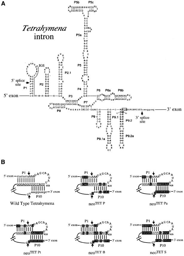
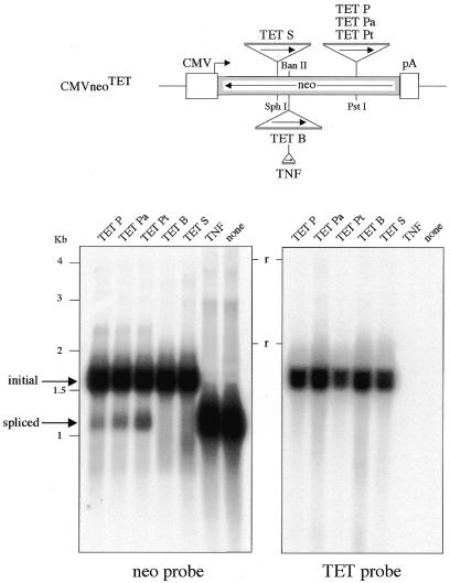
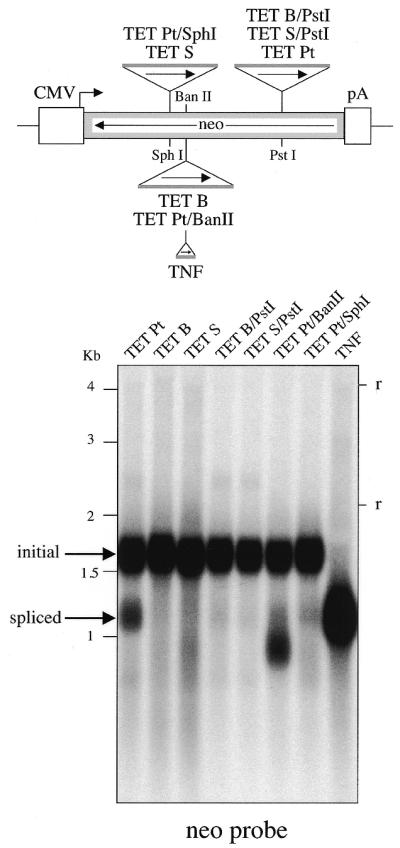
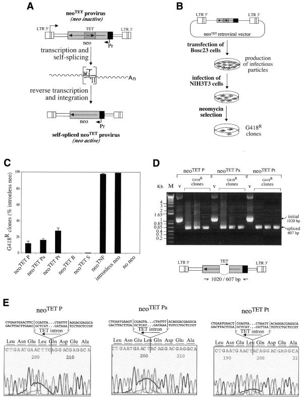
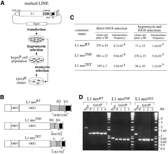
We devised an indicator gene for retrotransposition based on an autocatalytic ribozyme element--the Tetrahymena thermophila 23S rRNA group I intron--which can self-splice in vitro and does not require--at variance with nuclear mRNA introns--any specific pathway and cellular component for the completion of the splicing process. Several constructs, with the Tetrahymena intron adequately modified so as to be inserted at various positions within a neomycin-containing cassette under conditions that restore the neomycin-coding sequence after splicing out of the intron, were assayed for splicing efficiency in mammalian cells in culture. We show, both by northern blot analysis and by the recovery of neomycin activity upon retroviral transduction of the cassettes, that splicing efficiency depends on both the local base pairing and the global position of the intron within the neomycin transcript, and that some constructs are functional. We further show that they allow the efficient sorting out of retrotransposition events when assayed, as a control, with a human LINE retrotransposon. These indicator genes should be of great help in elucidating the mechanisms of transposition of a series of retroelements associated with transcripts not prone to nuclear mRNA intron splicing and previously not opened to any retrotransposition assay.


Figures

Figure 1
Figure 1
Structure of the wild-type T.thermophila rRNA group I intron and derivatives. (A) Predicted secondary structure of the Tetrahymena rRNA self-splicing intron with hairpins (Pi) schematized (according to 27). The 5′ and 3′ exonic sequences are in lower case and the intronic sequence in capital letters, with the splice sites indicated with arrows. The IGS (solid line) is essential for the splicing process and is involved in a complex association [see text and (B)]. (B) Schematic representation of the interactions involved in Tetrahymena intron self-splicing, with the pairing between the IGS and the 5′ and 3′ exonic sequences indicated with bars. The IGS sequence is underlined and the bulk of the intron sequence has been deleted. Mutations (indicated in light inverted) within the IGS and exonic sequences were generated (using appropriate primers and PCR amplification) to introduce the Tetrahymena intron into three distinct positions (the SphI, BanII and PstI sites) of the neomycin gene (see text and Fig. 2). The arrows delineate the 5′ and 3′ splice sites, and the exonic and intronic sequences are shown in lower and upper cases, respectively. The interactions of the IGS with the 5′ and 3′ exonic sequences (P1 and P10, respectively) are drawn simultaneously, for convenience.
Figure 2
Figure 2
Assay for in vivo splicing of the modified Tetrahymena introns, as followed by northern blot analysis. Top, schematic representation of the neomycin gene, with the positions (and orientation, see arrows) of the inserted Tetrahymena introns as modified and depicted in Figure 1. The neomycin gene (in reverse orientation, see arrow) is under the transcriptional control of the CMV promoter (CMV) and the SV40 polyadenylation signal (pA). Bottom, in vivo splicing of the intron-containing neo constructs. The intron-containing neo constructs were introduced by transfection into HeLa cells, and RNAs extracted 3 days post-transfection. Northern blot analysis was performed using total RNA (10 µg per lane) and hybridization with a neo probe (left) and a TET probe (right). Bands of 1.6 and 1.2 kb should be observed for the unspliced and spliced transcripts, respectively (arrows). Lanes P, Pa, Pt, B and S, modified Tetrahymena introns (see Fig. 1); lane TNF, nuclear mRNA intron 2 from the TNF β gene inserted at the BanII site (see scheme above and Materials and Methods); lane none, intronless neo construct. r, ribosomal RNAs.
Figure 3
Figure 3
Position effects for the in vivo splicing of the modified Tetrahymena introns. Same experimental conditions as in Figure 2, with the TET Pt, TET B and TET S introns inserted at their initial positions (lanes TET Pt, TET B and TET S) and at alternate positions (lanes TET B/PstI, TET S/PstI, TET Pt/BanII and TET Pt/SphI).
Figure 4
Figure 4
Quantitative assay for the splicing of the modified Tetrahymena introns and for the neoTET cassettes. (A) Rationale of the assay: in the initial copy of the neoTET marked gene, the neomycin gene-coding sequence (neo) is interrupted by the Tetrahymena intron, and cells containing the marked gene should be G418-sensitive. Upon transcription of the marked gene, the Tetrahymena intron should be spliced out and, after reverse transcription and integration (i.e. retrotransposition), the neo gene in the transposed copy should be active and the cells be G418-resistant. (B) Experimental procedures for the assay of the neoTET cassette. The neoTET cassette was introduced into a Moloney murine leukemia virus-derived vector, as illustrated. The corresponding plasmids were then introduced by transfection into a viral packaging cell line (Bosc23); the supernatants of the transfected packaging cells were collected, and used to infect target NIH3T3 cells. The 3T3 cells were then submitted to G418 selection to quantify the number of G418-resistant cells. DNAs from the resulting clones were finally extracted and analyzed (D and E) for the precise splicing out of the Tetrahymena intron. (C) Splicing and reporter gene efficiency as measured by the number of G418-resistant clones after retroviral transduction of the neo-containing cassettes. G418 selections were performed on plates seeded with 5 × 10⁴ cells (NIH3T3) per plate, and infected with 1 ml of transfected Bosc23 supernatant. The number of foci per plate is indicated for the five modified Tetrahymena intron-containing cassettes, the TNF intron-containing cassette, a positive control with an intronless neo cassette taken as a reference (100%) and a negative control without the neo gene; bars indicate standard deviations. (D) DNAs from individual clones were assayed by PCR for the presence of spliced copies, using primers bracketting the intronic domain. Positions of the primers used and sizes of the expected bands are indicated in the schema below the BET-labeled agarose gel. Bands of the expected size for the spliced-out intron (607 bp) are observed for all tested G418R clones, with a larger band (1020 bp) in the three control lanes (v, vector DNA). (E) Sequencing of the junctions of the spliced Tetrahymena introns. PCR products in (D) were sequenced, disclosing precise splicing out of the modified P, Pa and Pt Tetrahymena introns which restores the phase of the neo-coding sequence.
Figure 5
Figure 5
Quantitative assay for detection of retrotransposition of human LINEs marked with neo-cassettes. (A) Experimental procedures for detection of L1 retrotransposition events. The human L1.2 element (open box) was marked with a neo-cassette (neoTET, neoTNF or neoRT) and introduced into an hygromycin resistance gene-containing episomal vector. Marked L1-containing vectors were introduced into human cells by transfection, and then either cell transformants were isolated upon hygromycin selection and the resulting cell population (hygroR) was thereafter assayed for L1 retrotransposition upon G418 selection or, alternatively, transfected cells were directly submitted to G418 selection. (B) Structure of the three indicator genes (neoRT, neoTNF and neoTET) used for the L1 retrotransposition assay, with the RT and TNF introns being nuclear mRNA introns, and the Tetrahymena TET intron an autocatalytic group I intron. (C) Retrotransposition of the marked L1s. Numbers of foci per plates (5 × 10⁵ and 5 × 10⁴ cells per plate for direct and indirect G418 selections, respectively) are indicated as the mean of four independent transfections assays with the standard errors, together with the corresponding retrotransposition frequencies. (D) PCR analysis of the DNAs from initial and G418R cells assayed for the presence of retrotransposed copies with spliced-out introns, using the same primers bracketting the intronic domain for the three indicator genes. In the BET-labeled agarose gel, the first lane (hygR) for each construct corresponds to the DNA from the transfected hygroR cell population, before G418 selection, and the following lanes correspond to DNA from G418R cell populations (P) and clones (–4). Bands of the expected size for the spliced-out intron are observed only for the G418R cells. Larger bands, corresponding for each construct to unspliced copies, are observed in the initial hygroR cell populations, before G418 selection.

References

    1. Heidmann T., Heidmann,O. and Nicolas,J.-F. (1988) An indicator gene to demonstrate intracellular transposition of defective retroviruses. Proc. Natl Acad. Sci. USA, 85, 2219–2223. - PMC - PubMed
    1. Boeke J.D. and Stoye,J.P. (1997) Retrotransposons, endogenous retroviruses, and the evolution of retroelements. In Coffin,J.M., Hughes,S.H. and Varmus,H.E. (eds), Retroviruses. Cold Spring Harbor Laboratory Press, Cold Spring Harbor, NY, pp. 343–435. - PubMed
    1. Heidmann O. and Heidmann,T. (1991) Retrotransposition of a mouse IAP sequence tagged with an indicator gene. Cell, 64, 159–170. - PubMed
    1. Jensen S. and Heidmann,T. (1991) An indicator gene for detection of germline retrotransposition in transgenic Drosophila demonstrates RNA-mediated transposition of the LINE I element. EMBO J., 10, 1927–1937. - PMC - PubMed
    1. Moran J.V., Holmes,S.E., Nass,T.P., DeBerardinis,R.J., Boeke,J.D. and Kazazian,H.H.J. (1996) High frequency retroposition in cultured mammalian cells. Cell, 87, 917–927. - PubMed

Publication types

MeSH terms