. 2002 Jun 11;99(12):8173-8.
doi: 10.1073/pnas.122228699. Epub 2002 May 28.

Increased ionizing radiation sensitivity and genomic instability in the absence of histone H2AX

Affiliations

Increased ionizing radiation sensitivity and genomic instability in the absence of histone H2AX

Craig H Bassing et al. Proc Natl Acad Sci U S A. .

Abstract

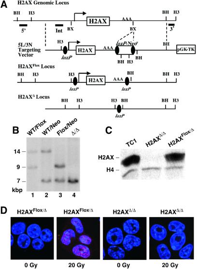
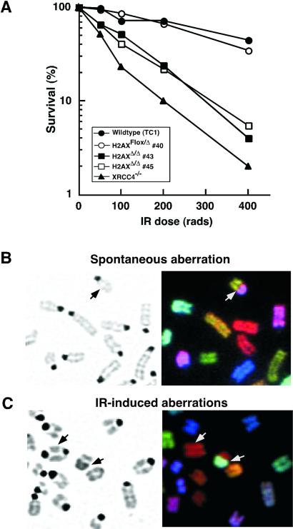
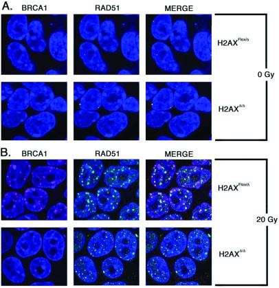
In mammalian cells, DNA double-strand breaks (DSBs) cause rapid phosphorylation of the H2AX core histone variant (to form gamma-H2AX) in megabase chromatin domains flanking sites of DNA damage. To investigate the role of H2AX in mammalian cells, we generated H2AX-deficient (H2AX(Delta)/Delta) mouse embryonic stem (ES) cells. H2AX(Delta)/Delta ES cells are viable. However, they are highly sensitive to ionizing radiation (IR) and exhibit elevated levels of spontaneous and IR-induced genomic instability. Notably, H2AX is not required for NHEJ per se because H2AX(Delta)/Delta ES cells support normal levels and fidelity of V(D)J recombination in transient assays and also support lymphocyte development in vivo. However, H2AX(Delta)/Delta ES cells exhibit altered IR-induced BRCA1 focus formation. Our findings indicate that H2AX function is essential for mammalian DNA repair and genomic stability.


Figures

Figure 1
Figure 1
Generation and characterization of H2AX-deficient ES Cells. (A) Schematic diagram of the mouse H2AX genomic locus, 5L/3N targeting vector, and H2AXFlox and H2AXΔ alleles. The H2AX promoter, exon, and polyadenylation site are shown, respectively, as an arrow, open box, and AAA. Filled bars show the relative locations of the 5′H2AX, IntH2AX, and 3′H2AX probes. Restriction site designations: BX, BstXI; BH, BamHI; H3, HindIII. (B) Southern blot analysis of H2AXWT/Neo (lane 2), H2AXWT/Flox (lane 1), H2AXFlox/Neo (lane 3), and H2AXΔ/Δ (lane 4) HindIII-digested DNA probed with 3′H2AX. The sizes of the bands are indicated. (C) Western blot analysis of TC1, H2AXFlox/Δ (no. 40), and H2AXΔ/Δ (no. 45) ES cells with anti-H2AX and anti-H4 antibodies (loading control). (D) Unirradiated and irradiated (15 min post 20 Gy) H2AXFlox/Δ and H2AXΔ/Δ cells immunostained for γ-H2AX foci (red).
Figure 2
Figure 2
Increased ionizing radiation sensitivity and genomic instability of H2AX-deficient ES cells. (A) IR sensitivity of wild-type (TC1), XRCC4−/−, H2AXFlox/Δ (no. 40), and two independent H2AXΔ/Δ (nos. 43 and 45) ES cell lines. Data are plotted as the percentage of colonies that grew out at a given γ-ray dose over unirradiated cells. The plotted numbers were obtained from triplicate data points of a representative experiment. (B and C) Metaphase spreads of unirradiated (B) and γ-irradiated (C) H2AXΔ/Δ ES cells. (Left) 4′,6-diamidino-2-phenylindole (DAPI)-stained chromosomes. (Right) SKY analysis. Arrow points to chromosomal translocations between two different chromosomes (B and C) and a dicentric chromosome (C).
Figure 3
Figure 3
Formation of IR-induced BRCA1 and RAD51 foci in H2AXΔ/Δ vs. H2AXFlox/Δ ES cells. Immunofluorescence of H2AXFlox/Δ and H2AXΔ/Δ cells unirradiated (A) or 6 h postirradiation with 20 Gy (B) to visualize foci of BRCA1 (rhodamine, red) and RAD51 (FITC, green). Nuclei appear blue (Topro-3). In the merged images, overlapping foci appear yellow.

References

    1. Wolffe A P. Chromatin Structure and Function. New York: Academic; 1998.
    1. Redon C, Pilch D, Rogakou E, Sedelnikova O, Newrock K, Bonner W. Curr Opin Genet Dev. 2002;12:162–169. - PubMed
    1. Abraham R T. Genes Dev. 2001;15:2177–2196. - PubMed
    1. Hoeijmakers J H. Nature (London) 2001;411:366–374. - PubMed
    1. Cromie G A, Connelly J C, Leach D R. Mol Cell. 2001;8:1163–1174. - PubMed

Publication types

MeSH terms