. 2002 May;14(5):1067-75.
doi: 10.1105/tpc.010475.

Photoexcited structure of a plant photoreceptor domain reveals a light-driven molecular switch

Affiliations

Photoexcited structure of a plant photoreceptor domain reveals a light-driven molecular switch

Sean Crosson et al. Plant Cell. 2002 May.

Abstract

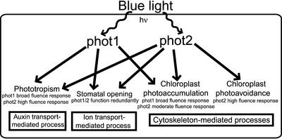
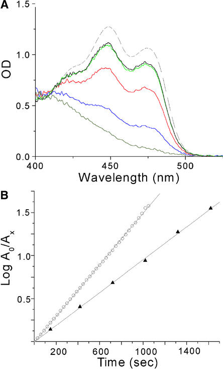
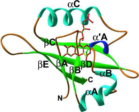
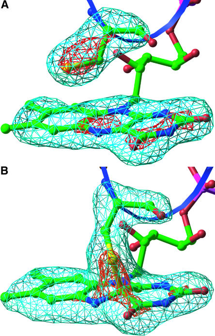
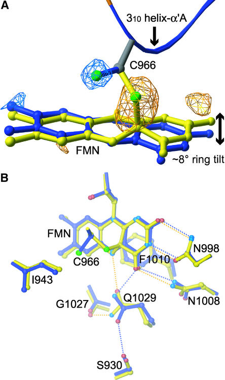
The phototropins are flavoprotein kinases that control phototropic bending, light-induced chloroplast movement, and stomatal opening in plants. Two flavin mononucleotide binding light, oxygen, or voltage (LOV) domains are the sites for initial photochemistry in these blue light photoreceptors. We have determined the steady state, photoexcited crystal structure of a flavin-bound LOV domain. The structure reveals a unique photochemical switch in the flavin binding pocket in which the absorption of light drives the formation of a reversible covalent bond between a highly conserved Cys residue and the flavin cofactor. This provides a molecular picture of a cysteinyl-flavin covalent adduct, the presumed signaling species that leads to phototropin kinase activation and subsequent signal transduction. We identify closely related LOV domains in two eubacterial proteins that suggests the light-induced conformational change evident in this structure is an ancient biomolecular response to light, arising before the appearance of plants.


Figures

Figure 1.
Figure 1.
Scheme of the Cellular Processes Affected by phot1 and phot2 and Their Fluence Dependence. The phot1 protein functions as a photoreceptor for phototropism and chloroplast photoaccumulation under a broad fluence range. phot2 functions as a photoreceptor for chloroplast photoaccumulation at moderate fluences and photoavoidance at high fluences. In addition, phot2 can function as a photoreceptor for phototropism under high fluence. Both phot1 and phot2 function as redundant photoreceptors that control stomatal opening. The initial photochemical step of cysteinyl-flavin adduct formation is identical in both of these plant blue light photoreceptors. The energy of a blue-light photon is designated hν.
Figure 2.
Figure 2.
Absorption Spectra and the Photocycle of phy3 LOV2 in the Crystal and in Solution. (A) Dark-state absorption spectra of phy3 LOV2 in single monoclinic crystals (solid black line) and solution (dashed line) normalized at 411 nm. Actinism of the monitoring light source contributes to a crystal dark-state spectrum containing a small percentage of photoproduct. Decay from the fully bleached spectrum of phy3 LOV2 (gray) is shown at 4 min (blue), 15 min (red), and 40 min (green). (B) Recovery of the dark state from the photoexcited steady state in solution (open circles) and in the crystal (closed triangles) monitored at 473 nm. A0 represents the amount of the nonadduct form of LOV2 at time 0, and Ax represents the amount at time x. The linearity of the plots indicates single exponential behavior with decay rates of 0.003 sec−1 in solution and 0.002 sec−1 in the crystal. This kinetic difference corresponds to a difference in the free energy of activation of 0.24 kcal/mol between LOV2 decay in the crystal and in solution.
Figure 3.
Figure 3.
Structure of Photoexcited phy3 LOV2. Ribbon diagram of the phy3 LOV2 structure under steady state illumination. The FMN cofactor is shown in the center of the fold with the bonds colored red. The sulfur of the conserved LOV Cys residue is pictured attached covalently to carbon 4a of FMN with a yellow adduct bond. Atoms of the Cys side chain and FMN are colored by elements: carbon, green; nitrogen, blue; phosphorus, pink, sulfur, yellow; and oxygen, red. The image was generated in Ribbons (Carson, 1997). Compare with Figure 1B from Crosson and Moffat (2001).
Figure 4.
Figure 4.
Light-Driven Cysteinyl-Flavin Adduct Formation in LOV2. Fourfold noncrystallographic symmetry–averaged omit maps of FMN and Cys-966 were calculated from the dark state (Crosson and Moffat, 2001) (A) and the photoexcited state (B) of phy3 LOV2. Maps are contoured at ±5σ and ±9σ, in which σ is the root-mean-square value of the electron density. The blue ribbon represents the 310 helix that contains the Cys. Atom colors are as in Figure 3. The images were generated in Ribbons (Carson, 1997).
Figure 5.
Figure 5.
Conformational Change in the FMN Binding Pocket of Photoexcited LOV2. (A) Fourfold noncrystallographic symmetry–averaged light-minus-dark difference Fourier map contoured at ±4σ, in which σ is the root-mean-square value of the electron density. Photoactive Cys and the flavin ring for the dark (blue) and photoexcited (yellow) structures are shown. Negative difference density (blue) and positive density (yellow) indicate Cys and ring motion upon photoexcitation. (B) Side chains exhibiting significant displacements between the dark (blue) and photoexcited (yellow) structures in response to cysteinyl-flavin C(4a) adduct formation. Hydrogen bonds between the protein and FMN cofactor in the dark and photoexcited structures are indicated by blue and yellow dotted lines, respectively. A 2.6 to 3.5 Å range for hydrogen bonding was used. Atoms are colored by elements: nitrogen, light blue; sulfur, green; and oxygen, red. Atoms colored blue in the dark structure and yellow in the photoexcited structure are carbon.
Figure 6.
Figure 6.
Alignment of LOV Domains with Known and Predicted Photoactivity. Phototropin and phy3 LOV domains with known photoactivity include Arabidopsis phot1 and phot2 and maidenhair fern (Adiantum) phy3. LOV domains from proteins that regulate circadian rhythm include Arabidopsis ZTL and FKF1 and Neurospora Wc-1 and VVD. Sequences for two eubacterial proteins containing a LOV domain with a flavin binding motif and conserved Cys also are shown: Bacillus YtvA and a putative sensor kinase from Caulobacter. Flavin-interacting residues are marked above the sequences (inverted triangles), as are residues whose side chains exhibit significant motion upon photoexcitation (+). Identical (black) and similar (gray) residues are highlighted at a 50% cutoff. The secondary structure of LOV2 is marked above the alignment: β-strand (arrows) and helix (helices).

References

    1. Akbar, S., Gaidenko, T.A., Kang, C.M., O'Reilly, M., Devine, K.M., and Price, C.W. (2001). New family of regulators in the environmental signaling pathway which activates the general stress transcription factor σ(B) of Bacillus subtilis. J. Bacteriol. 183, 1329–1338. - PMC - PubMed
    1. Altschul, S.F., Madden, T.L., Schaffer, A.A., Zhang, J., Zhang, Z., Miller, W., and Lipman, D.J. (1997). Gapped BLAST and PSI-BLAST: A new generation of protein database search programs. Nucleic Acids Res. 25, 3389–3402. - PMC - PubMed
    1. Arabidopsis Genome Initiative. (2000). Analysis of the genome sequence of the flowering plant Arabidopsis thaliana. Nature 408, 796–815. - PubMed
    1. Bailey, S. (1994). The CCP4 suite: Programs for protein crystallography. Acta Crystallogr. D 50, 760–763. - PubMed
    1. Bolognesi, M., Ghisla, S., and Incoccia, L. (1978). Crystal and molecular structure of 2 models of catalytic flavo(co)enzyme intermediates. Acta Crystallogr. B 34, 821–828.

Publication types

MeSH terms

LinkOut - more resources