. 2002 Jun 15;99(12):4466-74.
doi: 10.1182/blood.v99.12.4466.

Arp2/3 complex is required for actin polymerization during platelet shape change

Affiliations

Arp2/3 complex is required for actin polymerization during platelet shape change

Zhi Li et al. Blood. .

Abstract

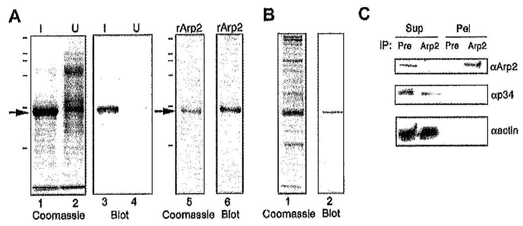
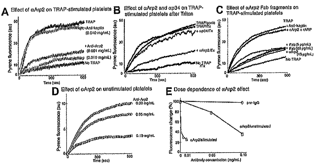
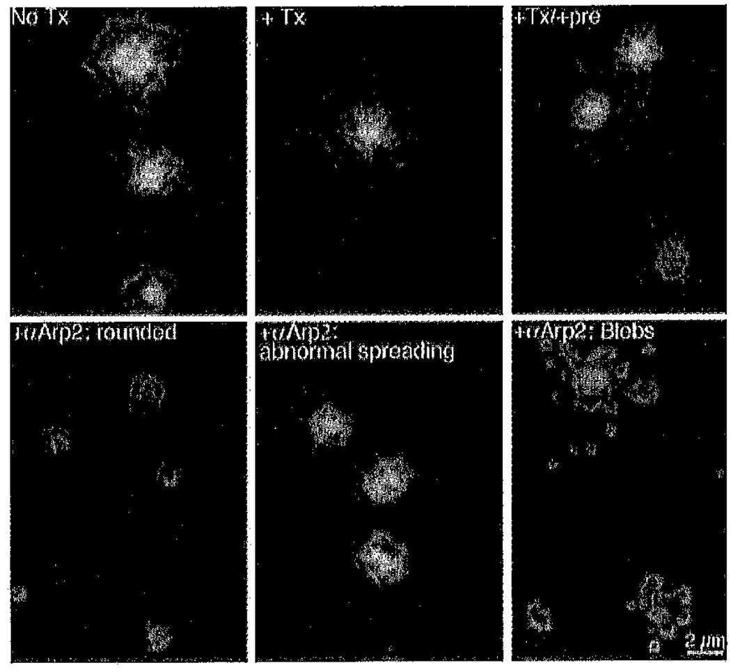
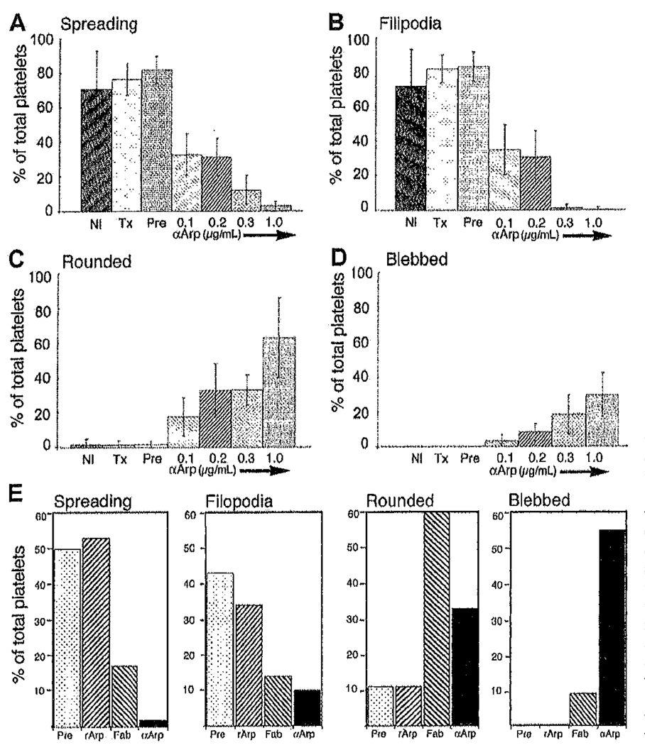
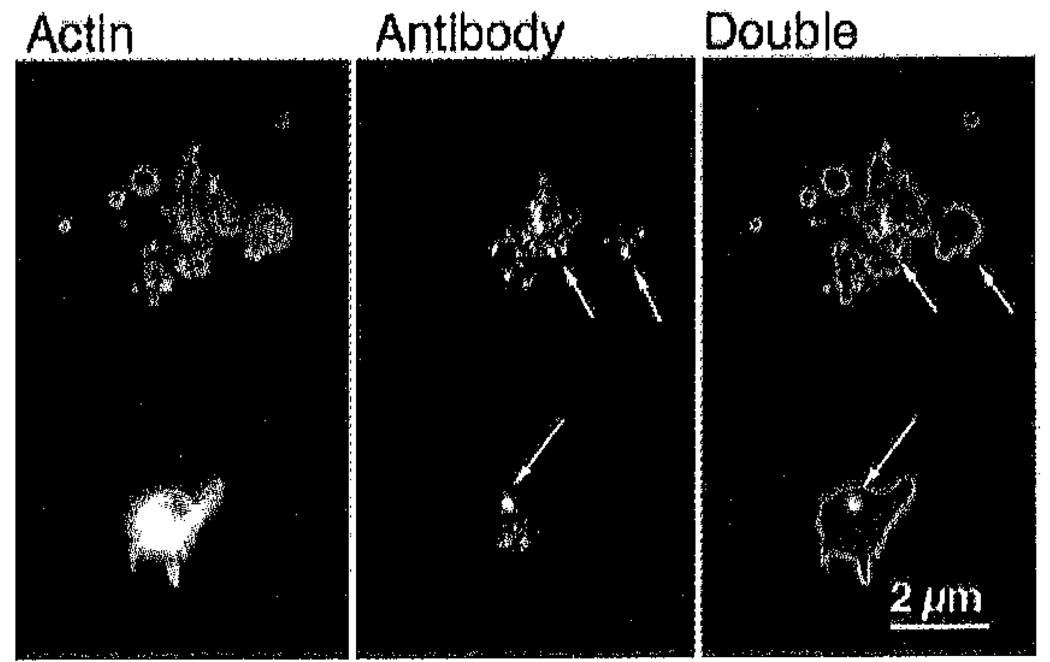
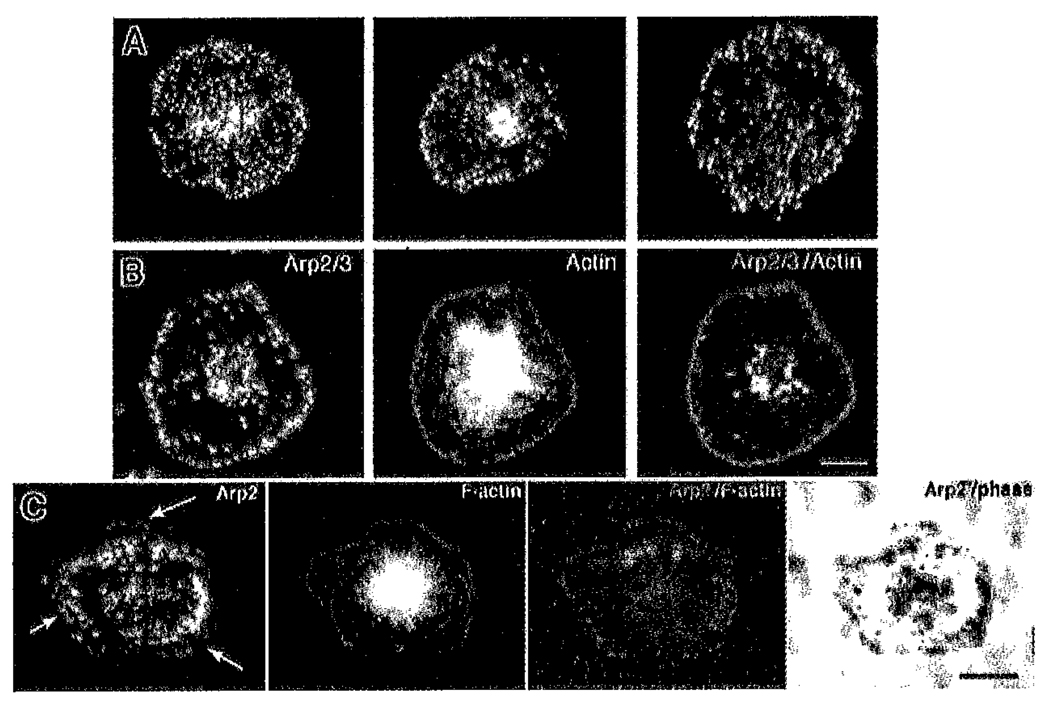
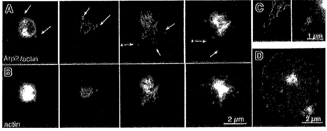
Platelets undergo a series of actin-dependent morphologic changes when activated by thrombin receptor activating peptide (TRAP) or when spreading on glass. Polymerization of actin results in the sequential formation of filopodia, lamellipodia, and stress fibers, but the molecular mechanisms regulating this polymerization are unknown. The Arp2/3 complex nucleates actin polymerization in vitro and could perform this function inside cells as well. To test whether Arp2/3 regulated platelet actin polymerization, we used recombinant Arp2 protein (rArp2) to generate Arp2-specific antibodies (alpha Arp2). Intact and Fab fragments of alpha Arp2 inhibited TRAP-stimulated actin-polymerizing activity in platelet extracts as measured by the pyrene assay. Inhibition was reversed by the addition of rArp2 protein. To test the effect of Arp2/3 inhibition on the formation of specific actin structures, we designed a new method to permeabilize resting platelets while preserving their ability to adhere and to form filopodia and lamellipodia on exposure to glass. Inhibition of Arp2/3 froze platelets at the rounded, early stage of activation, before the formation of filopodia and lamellipodia. By morphometric analysis, the proportion of platelets in the rounded stage rose from 2.85% in untreated to 63% after treatment with alpha Arp2. This effect was also seen with Fab fragments and was reversed by the addition of rArp2 protein. By immunofluorescence of platelets at various stages of spreading, the Arp2/3 complex was found in filopodia and lamellipodia. These results suggest that activation of the Arp2/3 complex at the cortex by TRAP stimulation initiates an explosive polymerization of actin filaments that is required for all subsequent actin-dependent events.


Figures

Figure 1
Figure 1. Characterization of αArp2 antibodies
(A) Affinity-purified αArp2 is specific for rArp2 in bacterial homogenates. Homogenates of IPTG-induced (I, lanes 1 and 3) or uninduced (U, lanes 2 and 4) M15 bacteria carrying the pQE30/Arp2 plasmid were electrophoresed in parallel. Gels were stained with Coomassie blue (lanes 1 and 2) or were Western blotted and probed with affinity-purified αArp2 antibodies (lanes 3 and 4). Coomassle blue-staining of purified recombinant Arp2 demonstrated a single band (lane 5) that was recognized by the affinity-purified antibody in Western blot (lane 6). In this case, 0.5 µg rArp2 has been loaded on the gel. (B) Detection of Arp2 in platelets by Western blot analysis. Platelet proteins were analyzed by Coomassie gels (lane 1) and for the presence of αArp2 in Western blots (lane 2). (C) Detection of native Arp2 in platelet extracts. Platelet extracts were incubated with protein A beads conjugated to either αArp2 (Arp2) or preimmune (Pre) antibodies from the same rabbit as αArp2, and the beads were collected by centrifugation. Supernatants (Sup) and pellets (Pel) were probed by Western blot for Arp2 (αArp2; top panel), p34, another Arp2/3 subunit (αp34; middle panel) or actin (αactin; lower panel). Note that Arp2 is removed from the extract and appears in the pellet after treatment with αArp2 beads but not with preimmune antibodies. Neither p34 nor actin sediments with the αArp2 beads.
Figure 2
Figure 2. αArp2 inhibits TRAP-stimulated actin-polymerizing activity
Platelets were stimulated with TRAP and were permeabilized with and without antibody as indicated. Actin-polymerizing activity was measured immediately after antibody addition. For each graph, all tracings are from the same preparations of platelets and actin preparation obtained on the same day. Results are presented in arbitrary units (au), as is standard in the field. Final fluorescence intensity with nonstimulated platelets (no TRAP) was defined as 10 au. Platelet extracts always produced at least a 20% increase over actin alone (not shown). (A) Effect of αArp2 on actin-polymerizing activity of TRAP-stimulated sonicated platelets. TRAP (top tracing) increased actin-polymerizing activity 5-fold over nonstimulated platelets (lowest tracing) in samples permeabilized by sonication. Affinity-purified αArp2 added to the TRAP-stimulated platelets decreased the initial rate and the extent of polymerization, whereas anti-kaptin had little to no effect at the same concentration (0.01 mg/mL, 0.7 µM) used for αArp2. (B) Effect of αArp2 and αp34 on actin-polymerizing activity of TRAP-stimulated Triton-permeabillzed platelets. Platelets were permeabilized by either sonication, as in Figure 1A, or with Triton and antibody added as indicated (αArp2 and αp34 were at 15 µg/mL [0.1 µM]). A similar increase in fluorescence was obtained with TRAP stimulation, whether platelets were permeabilized by sonication (sonic) or Triton (Tx). The increase in the initial rate of polymerization after TRAP stimulation was blocked by αArp2 regardless of whether permeabilization was with sonication or Triton. Less of an effect on TRAP-stimulated activity was detected with αp34. (C) Effect of Fab fragments of αArp2 on actin-polymerizing activity of TRAP-stimulated platelets. Platelets stimulated with TRAP and permeabilized as in Figure 1A were treated with antibodies as indicated at the following concentrations: anti-kaptin (0.1 µM), αArp2 + rArp2 (0.1 and 0.5 µM, respectively), Fab fragments of αArp2 (0.12 and 0.44 µM), and intact αArp2 from a different rabbit (0,1 µM), (D) Effect of αArp2 on actin-polymerizing activity of nonstimulated platelets. αArp2 was added (as indicated) to nonstimulated platelets immediately after sonication. Note that the arbitrary units (au) on the y-axis of the graph was expanded to increase the sensitivity to demonstrate the effect of the antibody on the lower level of activity present in nonstimulated platelets. (E) Dose dependence of αArp2 inhibition. The final fluorescence of representative experiments treated with increasing amounts of αArp2 or preimmune IgG (pre-IgG) is compared. A similar graph could be drawn for either anti-p34 or anti-kaptin as control antibody (Table 1). Experiments were normalized by setting the final fluorescence of parallel preparations, measured in the absence of antibody, at 100%, which allowed comparison of the low level of activity in nonstimulated platelets with the much higher activity after TRAP. Thus, results from TRAP-stimulated and nonstimulated experiments could be superimposed on the same graph.
Figure 3
Figure 3. αArp2 inhibits filopodia and lamellipodia formation during spreading on glass
Platelets were either directly spread on glass in plasma or were permeabilized briefly first with or without antibodies, and then mounted on glass, After 20 minutes, adherent platelets were fixed in the presence of FITC-phalloidin to stabilize and stain the F-actin. Top row of micrographs shows representative examples of spread platelets under 3 control conditions: no treatment (No Tx), permeabilized (+Tx), and permeabilized and loaded with preimmune antibodies (+Tx/+pre). The lower row of micrographs shows representative examples of platelets permeabilized and loaded with αArp2 before exposure to glass (+αArp2). Typical effects are frozen at the rounded stage (left panel), abnormal lamellipodia (middle panel), and blebbing (right panel).
Figure 4
Figure 4. Morphometric analysis of the effects of αArp2 on filopodia and lamellipodia and on morphology during spreading on glass
(A–D) Effect of Intact antibody on platelet spreading. Nl Indicates untreated (n = 259 platelets); Tx, permeabilized only (n = 391); Pre, permeabilized and loaded with 1.0 µg/mL antibodies from preimmune serum from the same rabbit (n = 444); and 0.1, 0.2, 0.3, and 1.0, permeabilized and loaded with different concentrations of αArp2, in µg/mL, final concentration (n = 423, n = 385, n = 512, n = 609, respectively). (E) Effect of Fab fragments and rArp2 on platelet spreading. Platelets were permeabilized as in Figure 3 and treated as follows: Pre indicates preimmune antibodies from the same rabbit (1 µg/mL); rArp2, αArp2 and recombinant Arp2 protein (1 µg/mL and 5 µg/mL, respectively); Fab, Fab fragments of αArp2 (1 µg/mL); and αArp2, intact αArp2 antibody (1 µg/mL). Note that Fab fragments freeze 60% of the platelets In the rounded stage, whereas pretreatment of the antibody with rArp2 protein eliminates this effect. Blebbing was seen with intact antibody and with Fab fragments, though less frequently with Fab fragments.
Figure 5
Figure 5. αArp2 antibody is detected in the cytoplasm after permeabilization
Representative examples of platelets permeabllized, loaded with αArp2, activated on glass, and fixed as in Figure 3. After fixation, platelets were stained with Cy3-labeled secondary antibody to determine whether the primary antibody gained access to the cytoplasm during permeabilization. Staining for αArp2 (red) and actin filaments with FITC–phalloidln (green) demonstrates diffuse speckling in the cytoplasm with some larger aggregates of antibody (arrows).
Figure 6
Figure 6. Location of endogenous Arp2/3 in spread platelets
(A) Three different spread platelets fixed in 4% paraformaldehyde containing 0.25% Triton and stained with αArp2. Arp2/3 displays a speckled cytoplasmic staining and a row of Immunofluorescent dots at the periphery, (B) Double label of a single platelet fixed as in panel A and stained for Arp2/3 with αp34 (Cy3, red) and for actin filaments (FITC–phalloidin, green). Superimposition of the double label shows Arp2/3 at the periphery. Bar = 2 µm. (C) Triple image of a single platelet fixed without detergent to permit imaging by phase microscopy and then stained with αArp2 (red) and phalloidin (green). Note that Arp2/3 is found in a bright arc internal to the actin frill. Arrows indicate weak staining at the outermost edge of the lamellipodia. Also note that the platelet contours imaged by phalloidin correspond to those imaged by phase microscopy. Bar = 2 µm.
Figure 7
Figure 7. Gallery of platelets at early stages of shape change showing localization of Arp2/3 at the cortex and at the tips and roots of filopodia
Representative double images of platelets fixed in the presence of detergent as in Figure 6A–B. (A) Four different platelets fixed at progressively later stages of activation from left to right and imaged for both F-actin (FITC–phalloidin, green) and Arp2/3 (Cy3, red). (B) Corresponding images of the same 4 platelets showing only the actin channel. (C) Higher magnification of the 2 filopodia indicated in panel A by an asterisk. (D) Fully spread platelet at the same magnification as the other platelets in this figure for size comparison.

References

    1. Escolar G, Krumwiede M, White JG. Organization of the actin cytoskeleton of resting and activated platelets in suspension. Am J Pathol. 1986;123:86–94. - PMC - PubMed
    1. Allen RD, Zacharski LR, Widirtsky ST, Rosenstein R, Zaitlin LM, Burgess DR. Transformation and motility of human platelets: details of the shape change and release reaction observed by optical and electron microscopy. J Cell Biol. 1979;83:126–142. - PMC - PubMed
    1. Hartwig JH. Mechanisms of actin rearrangements mediating platelet activation. J Cell Biol. 1992;118:1421–1442. - PMC - PubMed
    1. Bearer EL. Cytoskeletal domains in the activated platelet. Cell Motil Cytoskeleton. 1995;30:50–66. - PMC - PubMed
    1. Bearer EL, Abraham M. 2E4 (kaptin): a novel actin-associated protein from human blood platelets found in lamellipodia and the tips of the stereocilia of the inner ear. Eur J Cell Biol. 1999;78:117–126. - PMC - PubMed

Publication types

MeSH terms