

Review
doi: 10.1128/JCM.40.6.1892-1901.2002.
Laboratory identification of the microsporidia
Affiliations
- PMID: 12037040
- PMCID: PMC130667
- DOI: 10.1128/JCM.40.6.1892-1901.2002
Item in Clipboard
Review
Laboratory identification of the microsporidia
J Clin Microbiol.
2002 Jun.
No abstract available
Figures

Life cycle of microsporidia. Reprinted from reference
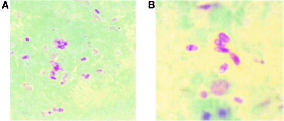
Weber-Green modified trichrome staining of microsporidial spores. (A) Spores in stool specimen; (B) higher magnification of the image shown in panel A.

Ryan-Blue modified trichrome staining of microsporidial spores. (A) Spores in stool specimen; (B) spores in intestinal tract tissue.

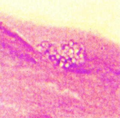
Single smear stained by an acid-fast trichrome stain method showing both an Isospora belli oocyst (modified acid-fast positive stain) and microsporidial spores (modified trichrome stain).

Giemsa staining of microsporidial spores in intestinal tract cells. The images show the development of the spores.
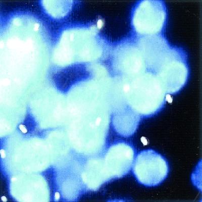
Calcofluor white staining of microsporidial spores in urine sediment.

Encephalitozoon spp. detected with immunofluorescent reagent. (A) Urine sediment; (B) positive control spores.

Cytospin preparation of bronchoalveolar lavage fluid from a patient with AIDS and intestinal E. bieneusi infection, showing intracellular gram-positive microsporidial spores (Gram stain). Reprinted from reference with permission.
Hematoxylin-eosin staining of eye tissue (note clear spores).

PAS staining of eye tissue (note PAS-positive granule at the end of each spore).

Warthin-Starry silver staining of eye tissue (note dark spores).

Transmission electron micrograph of a jejunal biopsy demonstrating numerous septated parasitophorous vacuoles of Encephalitozoon intestinalis, which are located in the Golgi-rich supranuclear cytoplasm. Reprinted from reference with permission.
References
-
- Canning, E. U. 1993. Microsporidia, p. 299-370. In J. P. Kreier (ed.), Parasitic Protozoa, vol. 6. Academic Press, San Diego, Calif.
-
- Croppo, G. P., G. S. Visvesvara, G. J. Leitch, S. Wallace, and D. A. Schwartz. 1998. Identification of the microsporidian Encephalitozoon hellem using immunoglobulin G monoclonal antibodies. Arch. Pathol. Lab. Med. 122:182-186. - PubMed
-
- Del Aguila, C., G. P. Croppo, H. Moura, A. J. Da Silva, G. J. Leitch, D. M. Moss, S. Wallace, S. B. Slemenda, N. J. Peiniazek, and G. S. Visvesvara. 1998. Ultrastructure, immunofluorescence, Western blot, and PCR analysis of eight isolates of Encephalitozoon (Septata) intestinalis established in culture from sputum and urine samples and duodenal aspirates of five patients with AIDS. J. Clin. Microbiol. 36:1201-1208. - PMC - PubMed
Publication types
MeSH terms
LinkOut - more resources
Full Text Sources
