

Detection of West Nile virus antigen in mosquitoes and avian tissues by a monoclonal antibody-based capture enzyme immunoassay
- PMID: 12037058
- PMCID: PMC130729
- DOI: 10.1128/JCM.40.6.2023-2030.2002
Detection of West Nile virus antigen in mosquitoes and avian tissues by a monoclonal antibody-based capture enzyme immunoassay
Abstract
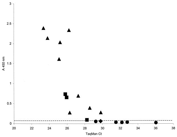
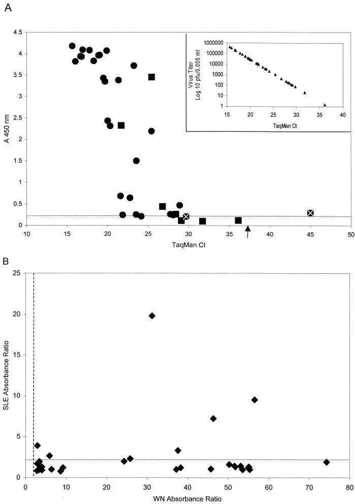
An antigen capture immunoassay to detect West Nile (WN) virus antigen in infected mosquitoes and avian tissues has been developed. With this assay purified WN virus was detected at a concentration of 32 pg/0.1 ml, and antigen in infected suckling mouse brain and laboratory-infected mosquito pools could be detected when the WN virus titer was 10(2.1) to 10(3.7) PFU/0.1 ml. In a blindly coded set of field-collected mosquito pools (n = 100), this assay detected WN virus antigen in 12 of 18 (66.7%) TaqMan-positive pools, whereas traditional reverse transcriptase PCR detected 10 of 18 (55.5%) positive pools. A sample set of 73 organ homogenates from naturally infected American crows was also examined by WN virus antigen capture immunoassay and TaqMan for the presence of WN virus. The antigen capture assay detected antigen in 30 of 34 (88.2%) TaqMan-positive tissues. Based upon a TaqMan-generated standard curve of infectious WN virus, the limit of detection in the antigen capture assay for avian tissue homogenates was approximately 10(3) PFU/0.1 ml. The recommended WN virus antigen capture protocol, which includes a capture assay followed by a confirmatory inhibition assay used to retest presumptive positive samples, could distinguish between the closely related WN and St. Louis encephalitis viruses in virus-infected mosquito pools and avian tissues. Therefore, this immunoassay demonstrates adequate sensitivity and specificity for surveillance of WN virus activity in mosquito vectors and avian hosts, and, in addition, it is easy to perform and relatively inexpensive compared with the TaqMan assay.
Figures
References
-
- Adams, S. C., A. K. Broom, L. M. Sammels, A. C. Hartnett, M. J. Howard, R. J. Coelen, J. S. Mackenzie, and R. A. Hall. 1995. Glycosylation and antigenic variation among Kunjin virus isolates. Virology 206:49-56. - PubMed
-
- Beaty, B. J., C. H. Calisher, and R. S. Shope. 1989. Arboviruses, p. 797-856. In N. J. Schmidt and R. W. Emmons (ed.), Diagnostic procedures for viral, rickettsial and chlamydia infections. American Public Health Association, Washington, D.C.
-
- Bowen, G. S., and D. B. Francy. 1980. Surveillance, p. 473-499. In T. P. Monath (ed.), St. Louis encephalitis. American Public Health Association, Washington, D.C.
-
- Brown, T. M., C. J. Mitchell, R. S. Nasci, G. C. Smith, and J. T. Roehrig. 2001. Detection of eastern equine encephalitis virus in infected mosquitoes using a monoclonal antibody based antigen-capture enzyme-linked immunosorbent assay. Am. J. Trop. Med. Hyg. 65:208-213. - PubMed
-
- Doggett, S. L., I. Koevski, J. Haniotis, and R. C. Russell. 1997. MOSAVEX: a mechanical device to grind mosquitoes for arbovirus detection. Arbovirus Res. Aust. 7:75-78.
Publication types
MeSH terms
Substances
LinkOut - more resources
Full Text Sources
Other Literature Sources
Medical
