. 2002 Jun;68(6):3076-84.
doi: 10.1128/AEM.68.6.3076-3084.2002.

Crescent bodies of Parachlamydia acanthamoeba and its life cycle within Acanthamoeba polyphaga: an electron micrograph study

Affiliations

Crescent bodies of Parachlamydia acanthamoeba and its life cycle within Acanthamoeba polyphaga: an electron micrograph study

Gilbert Greub et al. Appl Environ Microbiol. 2002 Jun.

Abstract

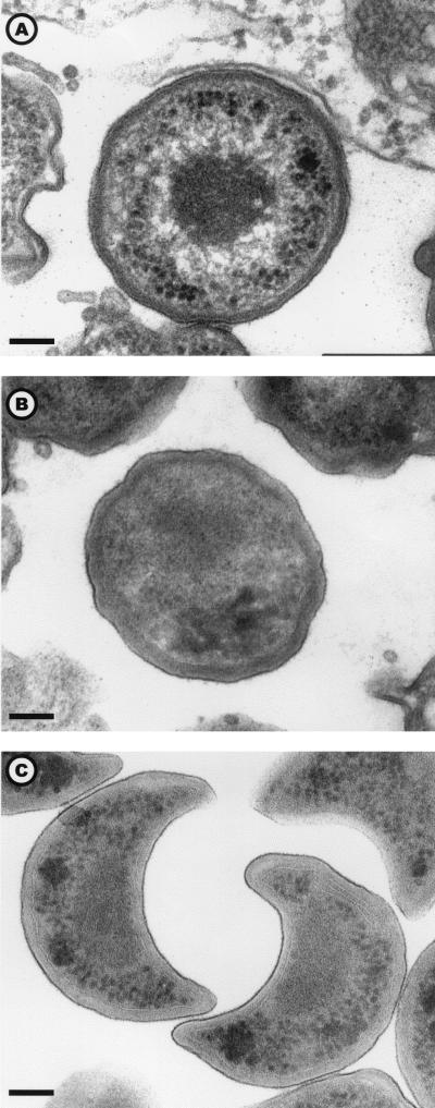
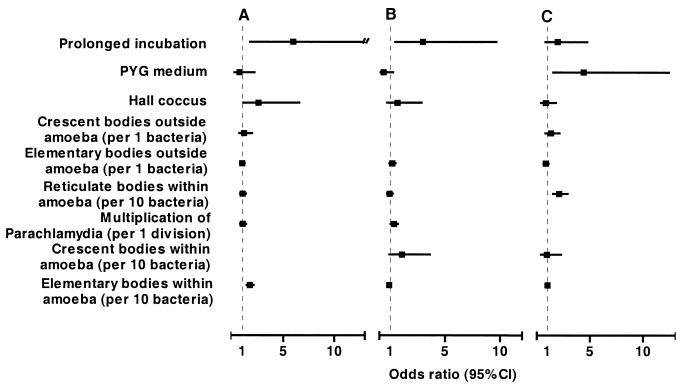
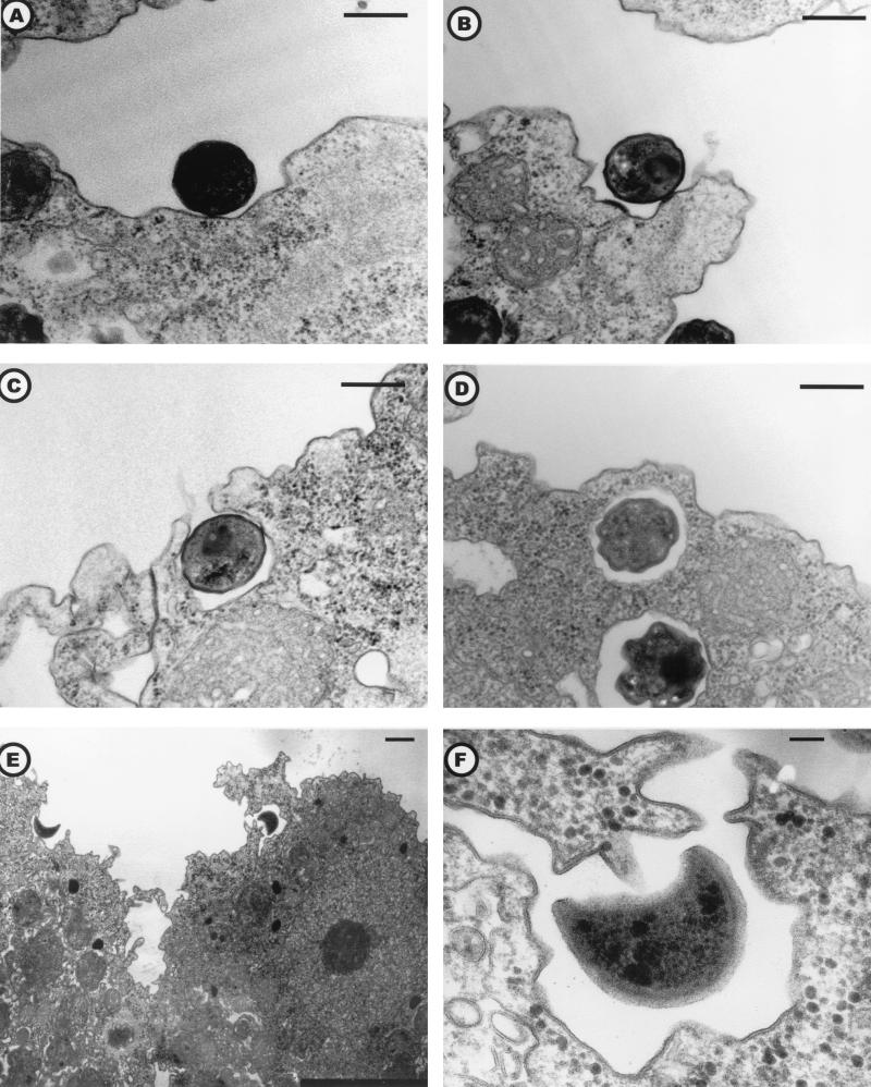
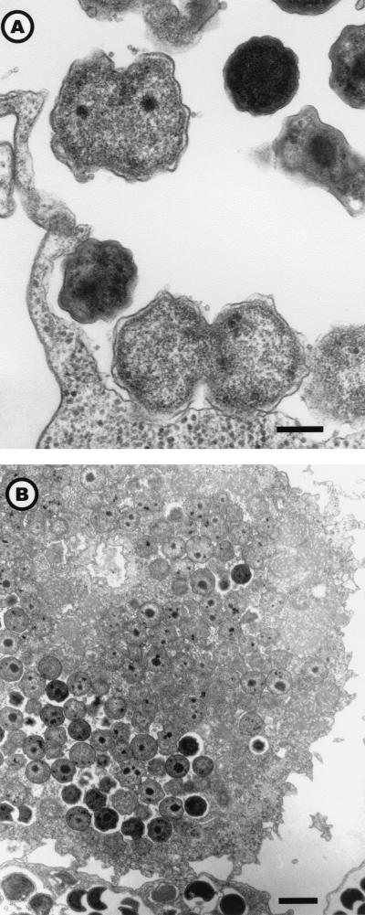
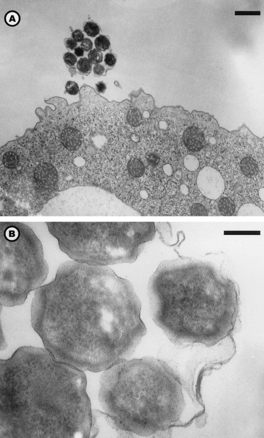
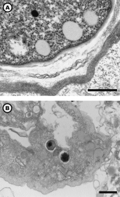
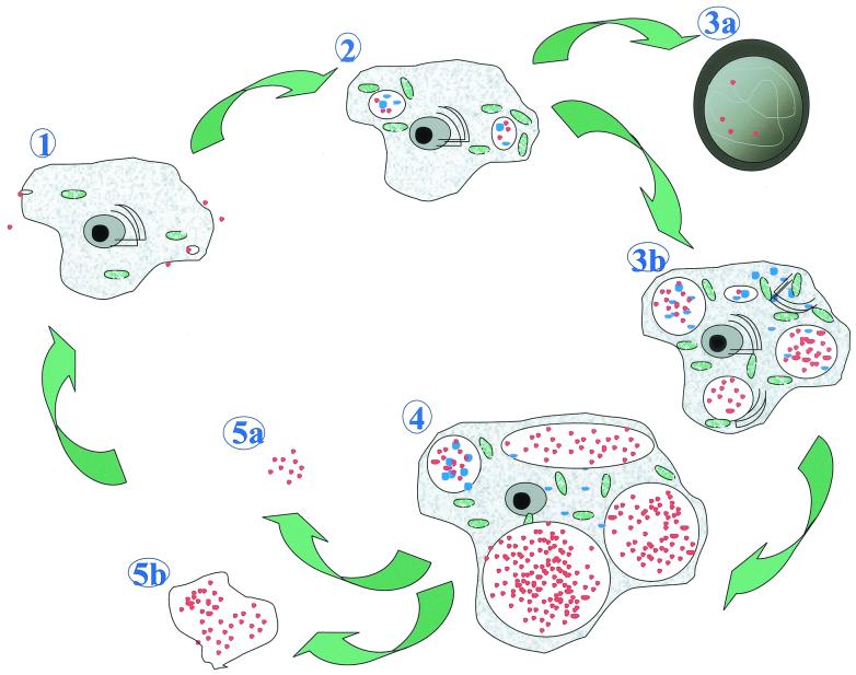
Parachlamydiaceae are endosymbionts of free-living amoeba first identified in 1997. Two developmental stages, elementary and reticulate bodies, were observed; however, their localization and proportions according to culture condition and duration remain unknown. The life cycle of Parachlamydia acanthamoeba within Acanthamoeba polyphaga was studied by transmission electron microscopy of 8-, 36-, and 144-h coculture. Morphometry and quantification were performed using SAMBA software. The elementary body, the predominant stage within the amoebae, was located mainly within their vacuoles. The multiplication of Parachlamydia bacteria by binary fission of reticulate bodies was independently associated with culture in PYG broth (odds ratio [OR] = 4.4; 95% confidence interval [CI], 1.55 to 12.46) and with the presence of reticulate bodies within the amoebae (OR = 2.10; 95% CI, 1.53 to 2.89). A third developmental stage was observed, the crescent body. Its presence outside and inside the amoebae was associated mainly with prolonged incubation time (OR = 3.98; 95% CI, 1.49 to 10.68, and OR = 5.98; 95% CI, 1.75 to 20.4, respectively). Elementary and crescent bodies were released into the extracellular medium within vesicles or after amoebal lysis. For both, phagocytosis was their mode of entry. This electron micrograph study revealed another infective developmental stage, the crescent body, and provided quantitative analysis of the life cycle of P. acanthamoeba within A. polyphaga.


Figures

FIG. 1.
FIG. 1.
The three developmental stages of Parachlamydia sp. Hall's coccus: the reticulate body (A), the elementary body (B), and the crescent body (C). Electron micrograph; magnification, ×45,600 (bar = 100 nm).
FIG. 2.
FIG. 2.
OR of having crescent bodies inside (A) and outside (B) A. polyphaga and of the presence of reticulate bodies undergoing binary fission (C). Data are adjusted for all variables listed.
FIG. 3.
FIG. 3.
Electron micrographs illustrating the phagocytosis of Hall's coccus elementary bodies (A to D) or BN9 endosymbiont crescent bodies (E and F). Magnifications: panels A to D, ×18,700 (bar = 500 nm); panel E, ×4,250 (bar = 1 μm); and panel F, ×51,000 (bar = 100 nm).
FIG. 4.
FIG. 4.
Binary fission (A) and invasion of the amoebal cytoplasm (B) by reticulate bodies. Hall’s coccus (A) and the BN9 endosymbiont (B) are shown. Electron micrograph; magnifications, ×18,400 (A) (bar = 300 nm) and ×4,600 (B) (bar = 1 μm).
FIG. 5.
FIG. 5.
Expelled vesicles filled with Parachlamydia sp. Hall's coccus. Electron micrograph; magnifications, ×5,000 magnification (bar = 1 μm) (A) and ×60,000 (bar = 100 nm) (B).
FIG. 6.
FIG. 6.
Uninfected A. polyphaga cyst (A) and infected amoeba in the process of being encysted (B). Electron micrograph; magnifications, ×30,240 (bar = 500 nm) (A) and ×9,240 (B) (bar = 1 μm).
FIG. 7.
FIG. 7.
Life cycle of P. acanthamoeba within A. polyphaga. (1) Entry of reticulate or crescent bodies (red dots) within the protozoa. (2) Differentiation in reticulate bodies (blue dots) and multiplication by binary fission within vacuoles. (3a) Encystment: trophozoites heavily infected by Parachlamydia lose their ability to encyst. (3b) Multiplication of the bacteria (many reticulate bodies, intravacuolar and intracytoplasmic), with increased vacuole and cytoplasm sizes. Dilatation of the endoplasmic reticulum (black semicircles) and metabolic activation (mitochondria, in green) are shown. (4) Amoeba with dilated vacuoles filled with elementary and crescent bodies. (5a) Free Parachlamydia bacteria. (5b) Expelled vesicles filled with Parachlamydia bacteria.

References

    1. Amann, R., N. Springer, W. Schönhuber, W. Ludwig, E. N. Schmid, K. D. Müller, and R. Michel. 1997. Obligate intracellular bacterial parasites of Acanthamoebae related to Chlamydia spp. Appl. Environ. Microbiol. 63:115-121. - PMC - PubMed
    1. Berk, S. G., R. S. Ting, G. W. Turner, and R. J. Ashburn. 1998. Production of respirable vesicles containing live Legionella pneumophila cells by two Acanthamoeba spp. Appl. Environ. Microbiol. 64:279-286. - PMC - PubMed
    1. Birtles, R. J., T. J. Rowbotham, D. Raoult, and T. G. Harrison. 1996. Phylogenetic diversity of intra-amoebal legionellae as revealed by 16S rRNA gene sequence comparison. Microbiology 142:3525-3530. - PubMed
    1. Birtles, R. J., T. J. Rowbotham, C. Storey, T. J. Marrie, and D. Raoult. 1997. Chlamydia-like obligate parasite of free-living amoebae. Lancet 349:925-926. - PubMed
    1. Borazjani, R. N., L. L. May, J. A. Noble, S. V. Avery, and D. G. Ahearn. 2000. Flow cytometry for determination of the efficacy of contact lens disinfecting solutions against Acanthamoeba spp. Appl. Environ. Microbiol 66:1057-1061. - PMC - PubMed

LinkOut - more resources